SonoStudy: Ultrasound for shoulder dislocation – Dx to anesthesia & reduction #FOAMed #FOAMus

A recent study in Annals of Emergency Medicine (found on pubmed too) discusses the use of ultrasound for assessing shoulder dislocation and reduction. Yup, that’s right – no need for that Xray – unless you are concerned about a fracture. But, when you have a patient with a history of shoulder dislocation saying, “it’s out again” then dont get that Xray – before or after your reduction – just use ultrasound. It’s quick and easy and can also be used for joint injections for anestheisa too. Dr. Mike Stone showed a great video of this too – 2 docs competing to see who finishes the assessment, anesthesia and reduction the quickest – guess who won….

Diagnostic Accuracy of Ultrasonographic Examination in the Management of Shoulder Dislocation in the Emergency Department

Study objective

Emergency physicians frequently encounter shoulder dislocation in their practice. The objective of this study is to assess the diagnostic accuracy of ultrasonography in detecting shoulder dislocation and confirming proper reduction in patients presenting to the emergency department (ED) with possible shoulder dislocation. We hypothesize that ultrasonography could be a reliable alternative for pre- and postradiographic evaluation of shoulder dislocation.

Methods

This was a prospective observational study. A convenience sample of patients suspected of having shoulder dislocation was enrolled in the study. Ultrasonography was performed before and after reduction procedure with a 7.5- to 10-MHz linear transducer. Shoulder dislocation was confirmed by taking radiographs in 3 routine views as a criterion standard. The operating characteristics of ultrasonography to detect dislocation in patients with possible shoulder dislocation and to confirm reduction in patients with definitive dislocation were calculated as the primary endpoints.

Results

Seventy-three patients were enrolled. The ultrasonography did not miss any dislocation. The results of ultrasonography and radiography were identical and the sensitivity of ultrasonography in detection of shoulder dislocation was 100% (95% confidence interval 93.4% to 100%). The sensitivity of ultrasonography for assessment of complete reduction of the shoulder joint reached 100% (95% confidence interval 93.2% to 100%) in our study as well.

Conclusion

We suggest that ultrasonography be performed in all patients who present to the ED with a clinical impression of shoulder dislocation on admission time. The results of this study provide promising preliminary support for the ability of ultrasonography to detect shoulder dislocation. However, further investigation is necessary to validate the results and assess the ability of ultrasonography in detecting fractures associated with dislocation.

To view Dr. Mike Stone’s lecture on shoulder dislocation diagnosed by ultrasound, view below:

For another great post of shoulder shrugging – see broomedocs site here!

ACEP News in 2/2014 had an article on shoulder dislocation by ultrasound – go here.

SonoStudy: Meta-analysis: History & Physical exam with Ultrasound for extremity fractures #FOAMed

I keep thinking about this study published in the Jan 2013 issue of Academic Emerg Med by Dr. Nikita Joshi et al…. for a few reasons… so I thought i would highlight it on SonoSpot and spark some discussion to get your thoughts too. (Get full article here). First off, it’s about a condition that I see in the emergency department on every shift, so it’s incredibly relevant. And, it involves imaging, specifically ultrasound, and how it can benefit the patients with this problem from cost savings to quicker diagnoses and treatment. Finally, the results actually surprised me. Not because ultrasound seemed to be just as good as radiographs, but that they weren’t better. But, I should say that it was a meta-analysis and quite difficult to compare and the study subjects in the meta-analysis all had radiograph proven fractures, and I wonder what would have happened if the xrays were negative but the bedside ultrasound was positive, proven by a gold standard, like CT scan???….  Who am I kidding?! That would involve too much cost, radiation, and time in the emergency department….. Oh wait, I get it….I guess I understand the importance of this study now. There have been quite a few studies on the topic in the last couple years – go here, here, here, here, and here – which makes it really exciting.

The authors start by stating that radiographs do miss fractures:

“The typical work-up of the injured patient generally involves a medical provider obtaining a history and physical examination, often followed by radiologic imaging. However, many times the radiologic imaging may be negative or inconclusive, which calls to question whether the imaging contributed to the management or outcome of the patient. Studies have shown that often the imaging obtained is unnecessary and results in radiation exposure to patients and increased ED wait times.[2]….There’s a low rate of positive radiography when assessing for fractures as evidenced by a retrospective review by Bentohami et al.,[3] in which only 50% of upper extremity x-rays showed fractures, and another study by Heyworth,[4] which showed 15% of patients with ankle injuries had documented fractures on x-ray. In the study by Stiell et al.,[2] patients with ankle injuries had midfoot fracture rates of 4.3%, and 9.3% had malleolar fractures. Therefore, 50% to 95% of extremity x-rays can be avoided without missing fractures.”

Ok, so we know this. Xrays arent great, so why get them? If you think the fracture would need reduction due to a displacement, then ok. But, wouldnt that be possible by physical exam as a deformed extremity so that you’d know to Xray that one? If the extremity is not deformed, but tender and swollen, why not just splint? Isnt that what you would do anyway if the xray was negative due to a high clinical concern for “occult fracture”?

The authors then follow this up with one of my favorite paragraphs on the topic:

“Bedside US has the potential benefits of reducing radiation exposure, costs, and pain, while potentially improving ED patient throughput and satisfaction. This reflects on the original purpose of developing CDRs for extremity fractures. Use of bedside US can help triage patients during a busy ED shift by quickly assessing for the presence of fracture as an adjunct to the normal history and physical examination. It can also aid nurses and physicians who may require more resources for reduction of a fracture.[11] EPs have become more adept at fracture diagnosis through independent review of US and radiographic imaging, and many researchers have examined the ability of EPs to obtain US imaging and diagnose fracture.[12, 13] Additionally, bedside US has excellent diagnostic test characteristics when performed by EPs compared to radiologists in the diagnostic evaluation for soft tissue infections,[14] cholecystitis,[15] pneumothorax,[16] or ruling out ectopic pregnancy.[17]

Love it. See the abstract below and read the entire article to see their limitations and methodology here.

Objectives

Understanding history, physical examination, and ultrasonography (US) to diagnose extremity fractures compared with radiography has potential benefits of decreasing radiation exposure, costs, and pain and improving emergency department (ED) resource management and triage time.

Methods

The authors performed two electronic searches using PubMed and EMBASE databases for studies published between 1965 to 2012 using a strategy based on the inclusion of any patient presenting with extremity injuries suspicious for fracture who had history and physical examination and a separate search for US performed by an emergency physician (EP) with subsequent radiography. The primary outcome was operating characteristics of ED history, physical examination, and US in diagnosing radiologically proven extremity fractures. The methodologic quality of the studies was assessed using the quality assessment of studies of diagnostic accuracy tool (QUADAS-2).

Results

Nine studies met the inclusion criteria for history and physical examination, while eight studies met the inclusion criteria for US. There was significant heterogeneity in the studies that prevented data pooling. Data were organized into subgroups based on anatomic fracture locations, but heterogeneity within the subgroups also prevented data pooling. The prevalence of fracture varied among the studies from 22% to 70%. Upper extremity physical examination tests have positive likelihood ratios (LRs) ranging from 1.2 to infinity and negative LRs ranging from 0 to 0.8. US sensitivities varied between 85% and 100%, specificities varied between 73% and 100%, positive LRs varied between 3.2 and 56.1, and negative LRs varied between 0 and 0.2.

Conclusions

Compared with radiography, EP US is an accurate diagnostic test to rule in or rule out extremity fractures. The diagnostic accuracy for history and physical examination are inconclusive. Future research is needed to understand the accuracy of ED US when combined with history and physical examination for upper and lower extremity fractures.

Nice job Nikita!

UltrasoundPodcast recently did a podcast on Distal radius fractures.

A great video of distal radius fractures can be seen here:

In case you’re curious about how easy it is to visualize a fracture by ultrasound, see image below. That bright white line is bone, and that break is …a break.

Screen Shot 2013-06-06 at 4.54.00 PM

SonoStudies: US for hip dislocations, septic hips, and fascia ilaca block for hip fractures

Quite a few recent studies on bedside ultrasound have focused on the hip, as it should, since it is so darn hard to evaluate it by the physical exam alone. Well, not only could ultrasound be used for diagnosing hip dislocations (as evidenced by the below case report), but it is also great for evaluating septic hip joints by visualizing the effusions and helping in its arthrocentesis needs …..as well as using ultrasound for ultrasound-guided fascia iliaca compartment block for hip fractures (especially in the elderly who you’d rather not give a ton of opiates to). – These are all from the Journal of EM.

The first case report discusses a 51 yr old man who was brought in the ED 20 minutes after a fall on wet grass while playing basketball (I know -good for him for staying active!). The current standard of care is to order an Xray. But, sometimes the Xray will not give you the information you need and you may go to CT, or the radiology tech is busy with traumas or other inpatient needs. In this case, the Ap Pelvis XR was normal. Well, never fear – the ultrasound is here! The diagnosis was made of an anterior hip dislocation by ultrasound. See the image below of his dislocated Right hip and normal Left hip when they used their curvilinear probe in anterior, mid-axial and coronal planes:

Screen shot 2012-12-18 at 7.28.25 PM

Another case report recently published discusses an 18 yr old female c/o 5 days of hip pain radiating down her anterior thigh and worse with weight bearing and hip movement. No fevers or other symptoms… oh, and she is 23 weeks pregnant. Now, the diagnosis of septic hips is a clinical one yet, sometimes, it can fool the best of us. I know Ive seen a patient with a septic hip walk…yes, with a limp, but still walk… saying “I think I just twisted it.” She was a bounce back to the (different) ED for persistent hip pain after an US was negative for DVT. She had mild leukocytosis (but what pregnant patient doesn’t!?!). The ED docs took a look with their ultrasound machine and saw an effusion (top picture below) (compared it to the opposite a-symptomatic hip (bottom picture below)) and then performed an ultrasound-guided arthrocentesis of purulent fluid: arrow and closed arrow is the femoral head and neck, respectively.

Screen shot 2012-12-18 at 7.48.32 PM

Screen shot 2012-12-18 at 7.49.41 PM

For a great podcast, the only way they know how to make it even more enjoyable – check out UltrasoundPodcast insert for hip ultrasund, aspiration and injection.

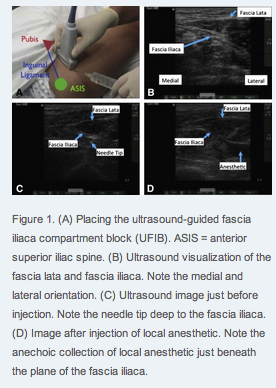
Now, the last study I will highlight, is one that is a more common concept/indication for hip issues – the fascia iliaca block for hip fractures. “”Hip fracture (HFx) is a painful injury that is commonly seen in the emergency department (ED). Patients who experience pain from HFx are often treated with intravenous opiates, which may cause deleterious side effects, particularly in elderly patients. An alternative to systemic opioid analgesia involves peripheral nerve blockade”  – word! A small study showing a decrease in pain scale in over 75% of the patients:

Screen shot 2012-12-18 at 7.56.45 PM

SonoArticle: Ultrasound FIRST for cellulitis/abscess evaluation – by Adhikari & Blaivas, oh yeah!

In a prior post we discussed the concept of what is now one of the post popular phrases that have come to be used as a Sono-term: ‘pus-stalsis’. Yup, that’s right  – the movement of pus seen with compression over the area of hypoechogenicity when using the linear probe to evaluate for fluid filled pocket that’s concerning for abscess. It can fool you! Instead of doing a needle aspiration – take a look! push down on it and see if there is pus-stalsis! It’s easy. Continue reading

SonoApp & Study: Cellulitis vs Abscess; US more sensitive than CT for soft tissue abscess

A patient comes into your emergency department or outpatient clinic that has a painful red area on their skin:

 -from Medicineo blog

…and you wonder whether its a superficial cellulitis, or if it’s a pus-filled abscess – and if it is an abscess, then how deep is it? how long is it? how loculated is it? Continue reading