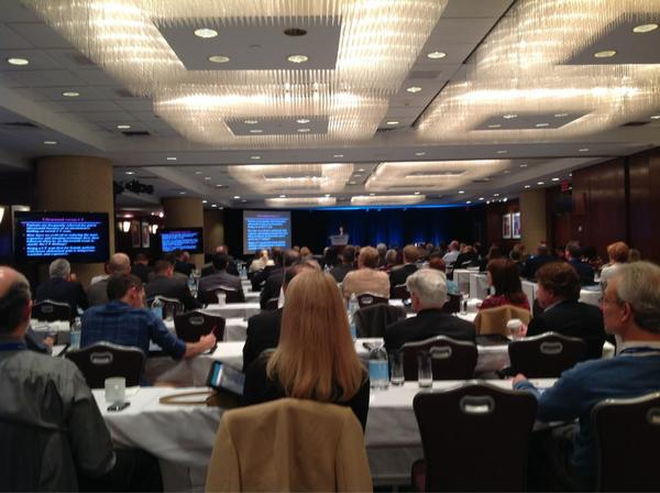
SonoWorkshop: CastleFest2014 #CF2014 – happening now, watch LIVE.. or after it’s done #FOAMed

The incredible team of Castlefest, has its 2014 sessions underway. The gurus are out in full force – Mike Mallin, Matt Dawson (@ultrasoundpod), Mike Stone (@bedsidesono), Chris Fox (@jchristianfox), Vicki Noble (@nobleultrasound), Haney Mallemat,(@CriticalCareNow), Rob Rogers (@EM_Educator), and many more!

What is Castlefest? Watch here to find out!

Watch the broadcast LIVE!

You can also take a look at the previous days’ lectures too: go here!

Day 1 Session 1 and Session 2

To find out more about the virtual ultrasound fellowship – the Ultrasound Leadership Academy – go here

SonoEvents: SonoGames, Castlefest2013, and the FREE Intro to Bedside Ultrasound iBook #FOAMed

This SonoGift is amazing! I could have sworn that I sent this earlier (and I think i did on Twitter and Facebook), but for whatever reason, it is in my blog’s draft folder, and I was shocked! – How dare I keep this away from everyone who follows SonoSpot?!!!! So, I apologize… from the bottom of my subxiphoid window (…ha! yes i know, I have many of them). If you’re getting this for the second time, then take it as a friendly reminder of how awesome the UltrasoundPodcast guys are to provide this amazing gift to everyone to learn the up-to-date info on bedside ultrasound applications… for free. Now, if you are getting this for the first time, you are going to LOVE it….. Why? Well, first off, it’s free (did I say that already?). And, if that wasnt enough, it’s the pdf version of the AWESOME iPAD download-able iBook (also found here chapter by chapter purchasing on inkling that can also been read on iPHONE) of Introduction to Bedside Ultrasound from the UltrasoundPodcast crew, with chapters written by so many of my friends. If you purchase the iPAD version (for pretty cheap, if you ask me) (including the iPAD mini and retina display), you can view all the clips and videos placed by the authors/experts in bedside ultrasound – which truly makes it the best “book” on bedside ultrasound that I know. It’s worth it.

You know what else Im excited about? SonoGames Part Deux at SAEM – this time, our crew is going to make it after the first round!!! You all better watch out! A sneak peak at SonoGames last year by the serious yet humorous, heavy yet light, good yet talented UltrasoundPodcast and their hilarious interviews:

Oh, and did I mention Castlefest2013????!! Im so excited to join them at CastleFest2013 – ultrasound, castles, wine, and festivities?—what more could a Sonogirl ask for?! You can even be there virtually! Yes, they did think of everything.

SonoWorkshop: Pearls (and more!) from the Stanford CME Ultrasound Course #FOAMed

Once again, our Stanford Ultrasound Workshop was a huge success. Why? Our instructors were phenomenal and from different specialties! Our participants were faculty from emergency medicine, internal medicine, critical care, surgery, and pediatrics! The ultrasound tips and tricks just kept on coming from our lecturers  – and, everyone laughed at our jokes, which always makes things great. As always, I like to provide those tips and tricks to all of you (and maybe even some of the jokes), so that you can feel like you were there too!

IMG_1087

Dr. Sarah Williams – First, the coordinator for the Stanford CME workshop welcomes everyone with a Star Wars phrase “Learning you are….May the force be with you, young padawans!” – always goes to a great start. She is also the creator of the Stanford Ultrasound Program and current Associate Residency Director (and the person who was kind enough to put up with my quirks and jokes to hire me as a fellow years ago). Her pearls on the EFAST: detects >600 cc (intraperitoneal) fluid, look around inferior pole in RUQ and subdiaphragm area of LUQ (free fluid develops first there!), it’s not good for pelvic fx/injuries (pelvic bleeding into pelvic cavity, and retroperitoneal, bowel gas obstructs view, bladder may be empty limiting visualization), it’s not done fast- FAST is part of RUSH, but dont rush the FAST. Look for your kidney, then look above it, around it and below it (thoracic fluid, morison’s pouch, paracolic gutter). FAST LUQ: higher, spleen smaller, stomach big -place knuckles on gurney, oblique probe in plane to ribs, free fluid can be between diaphragm & spleen.  #ultrasound detects 15-20cc fluid in thoracic cavity, better than chest Xray. Have patient take deep breath to lower diaphragm. The longer the patient is supine (or trendelenberg) the better, so if you have a walk-in trauma, perform serial FAST scan. SX view: the liver is the heart’s protector, be sure to see it in view- it allows you to see the 4chambers. gas is heart’s enemy – if gas gets in the way, you cannot see the liver: slide probe laterally to patient’s right, get that liver in your view. For pneumothorax eval – use linear probe, find your ribs, ID pleur liine, decr gain (brightness) to see sliding better. Start high in midclav line, indicator to head – – then travel thru mult rib spaces to estimate size.

IMG_1089

Dr. Laleh Gharahbaghian (since i cannot speak about myself, I let someone else write this part and promised I wouldn’t change anything – let us pray…): “With her usual stylish self, walking all around the workshop, giving hi-fives to everyone in her path, her dance moves came in handy as she spoke (can you point her out in this video from the mid 1990s of her past job?)  – She is the current Director of the Stanford Ultrasound Program and Fellowship her pearls can be found below: Her pearls of Aorta US: use large footprint probe, if get gas, press down, takes time – as if you were reducing a hernia; start in the subxiphoid region, travel down thru to iliacs. Most AAA are infrarenal and may seem normal in size at sx and get large once you travel down. Doesnt evaluate for rupture – most AAA leak/rupture retroperitoneal – not detected by US (your FAST is neg) – correlate clinically to your patient symptoms and vital signs. Her pearls on Renal US: main indication: hydronpehrosis, but pay attention to everything (outside to inside); eval both kidneys AND bladder- without bladder, you wont know if the bilateral hydro may just be that they have to pee. If empty bladder, and bilateral hydro, then possible mass (if not chronic). If patient is >50yo with flank pain, dont forget to eval the aorta as well. Start outside to inside for pathology – free fluid around kidney, cyst from kidney, mass on kidney, stone within kidney, hydronephrosis. Her pearls on Gallbladder US: start in the subxiphoid region, indicator to patient’s right, use liver as window, fan thru it medial to lateral to find GB. Then, fan/eval in transverse & longitudinal planes. Fanning thru the GB is key- there’ll be sections where it looks normal, then you fan & a stone comes into view! See if the stone is mobile by turning patient and re-scanning to see if moved. Think of the number 4 (or multiples of it) with measurements: width 4cm, length 8-10cm, anterior GB wall <4mm, CBD 4mm at 40yrs old (adding 1mm for every decade beyond).

IMG_1088

Dr. Phil Perera – our newest addition to the Stanford US team serving as the Director of US Research and the Associate Program Director gave quite an engaging talk on Echo and the RUSH protocol, putting it to action! As is highlighted by his Soundbytes website that is a free source of lectures for your viewing pleasure, he would keep asking the audience whether they would involve their consultants, if they would “write home to mom about this?!” The funny part is that one of the audience members responded with “I wouldn’t have to, she would call me before I get a chance.” Another participant concluded the point by saying, “You must work in a profitable community hospital.” Ah – gotta love the sarcasm! Phil continued his talk discussing when you should act fast by going through RUSH cases, giving props to others who also study and educate on resuscitation ultrasound. His pearls on EchoPSL view is the favorite – lets you evaluate right ventricle size, left ventricle size and contractility, pericardial effusion, pleural effusion and mitral valve regurge; Echo should be done with IVC when thinking about fluid resuscitation – if hyperdynamic -can tolerate fluid; if hypocontractile, not so much; Echo can eval aorta too! PSL view visualizes ascending aorta and descending aorta; AP4 view shows descending aorta – look for aneurysm/flap. Intraperitoneal fluid and pleural effusion can be mistaken for pericardial effusion – know where your pericardium is! Pleual effusion in PSL view travels behind descending aorta; pericardial effusion travels in front of descending aorta. AP4 great for comparing RV and LV chamber size, contractility of RV and LV. To get the P4 view, slide lateral after parasternal views until get to apex, angle to body center. His pearls on RUSH: Case that inspired him: 67yo acute SOB, in shock h/o COPD/CHF/HTN, CXR neg, ultrasound showing the cause to not be sepsis, but cardiogenic shock. RUSH provides the answer to : sepsis? cardiogenic? hypovolemic? hypervolemic? tamponade? PE? trauma? tension ptx? AAA? First & most important is the cardiac echo: the PUMP, that’s why it’s first – lots of info from a single cardiac view (PSL). For semi-quantitative contractility eval: fractional shortening & EPSS are measured – PSL must be at approp long section.  tamponade on #ultrasound – RV collapse during when it should fill (diastole)-also can see RA scalloping -do pericardiocentesis. pericardiocentesis: US studies show having pt in left lateral decubitus position & an apical view better for removing pericardial effusion than traditional SX technique. IVC – can use M mode to measure in both transv and long view 2cm from RA – can use your internal jugular as an alternate. Lung ultrasound – B Lines – think of fluids and your resuscitation when evaluating etiology of shock: FALLS protocol by Lichtenstein. Although rare, if your EKG has STEMI, do an ECHO – make sure its not a dissection before you start heparin!

IMG_1091 and Screen Shot 2013-03-01 at 1.41.03 PM

Dr. Zoe Howard (our Director of Medical Student and Resident US Education) and Dr. John Kugler (coordinator for the internal medicine US elective and global health US instructor) spoke about the many awesome ways ultrasound can help with procedural guidance. The dynamic duo had awesome videos to assist in their lecture and went through the many procedures that can be done with US guidance. Their pearls on Procedural US: On central line access – first look for the vein before you prep the area; it’s possible that the vein you want (or the location of the vein you want) is not the best vein for the procedure. Your indicator should be to your left, the screen dot should be on your left, that way left means LEFT when you’re guiding your needle tip to the vein. On lumbar puncture – do it when you can’t feel the landmarks, when you only have one attempt, when you’ve already had one unsuccessful attempt. Use the ALiEM trick with a paperclip for drawing the straight line. On thoracentesis and paracentesis – make sure you view the area where there is at least 2cm of fluid between the probe and the lung/bowel to avoid lung/bowel puncture – it may not always be where you think. On pericardiocentesis – look for where the fluid is most, patient to left lateral decubitus position, and you’ll find that SX is not the best anymore. On nerve blockslearn it, do it, and teach it! Your patients deserve it! Use the in-plane approach to visualize your entire needle, and use the dental syringe holder to have control over your syringe.

IMG_1093 Screen Shot 2013-03-01 at 2.05.40 PM Screen Shot 2013-03-01 at 2.03.42 PM Screen Shot 2013-03-01 at 2.04.57 PM Screen Shot 2013-03-01 at 2.06.47 PM Screen Shot 2013-03-01 at 2.59.35 PM Screen Shot 2013-03-01 at 3.07.32 PM

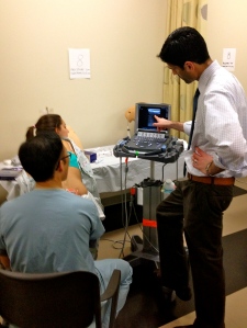
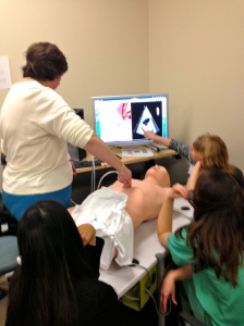
We then had 8 different hands-on stations with 11 different instructors (and 4 chickens used for central lines deserving of props, and lots of other simulators as you will see in the below pics)!  Including those described above, we also had the above instructors (from left to right, top to bottom (hyperlinks take you to “other” images that come up when you google search their names)- Dr. Viveta Lobo – our current US fellow and future Director of the Visiting Scholars Program, Dr. Brita Zaia – our past US fellow and current Kaiser San Francisco Ultasound Director, Dr. Darrel Sutijono – US fellowship trained faculty at Kaiser Santa Clara and new to twitter and #FOAMed and the FOAM movement, Dr. Manish Asarvala – US fellowship trained at UCSF and faculty at Kaiser Santa Clara. Dr. Yoshi Mitarai – an emergency medicine/critical care specialist who recently saved a life while at the gym doing Zumba (yup, you read that right), Dr. Suzanne Lippert – a specialist in nerve blocks and international/global health who is faculty at Stanford EM. Dr. Jennifer Newberry – an MD JD (so, super smart) and one of our senior EM residents who is staying on as a fellow in healthcare/ public policy.

IMG_1094IMG_1095 IMG_1097 IMG_1099 IMG_1100 IMG_1102 IMG_1103 IMG_1105

SonoMedStudent and UltraFest: Ultrasound in the Medical Schools…it’s time.

I just got back from the 2nd Annual UCI UltraFest – a FREE (yup, you read that right, FREE!) medical student ultrasound workshop held at UC Irvine this year for any and all medical students in California who want to come and learn bedside ultrasound using simulation and live models along with hearing about the future and international ultrasound. It’s getting a lot of press – as it should. It started off with an idea that came by one of my friends and mentors, Dr. Chris Fox (an ultrasound guru in his own right), and with the help of his medical students (Lancelot Beier, Kiah Bertoglio, and MaryJane Vennat), they organized faculty from all over California (UCI, Stanford, UCLA, USC) from multiple specialties. As Chris states “If I can give medical students the confidence and curiosity to want to use ultrasound all the time, then I feel they will take better care of their patients, and will provide more accurate care without turning to radiation.” You know how many medical students showed up? about 300. On a Saturday. During their free time. Some even wore a tie! …even when they ate from the free taco truck at lunch (yum!).

IMG_1084IMG_1077UltrafestpicUltrafestpic2ultrafestpic3For even more pics go here.

You know what that means? They want to learn it …and it should be incorporated into all medical school curriculums. It has been well studied to improve their learning in anatomy, physiology, and pathology… not to mention their patient care in their clinical years. It’s time. I’m excited to host it at Stanford for 2014!

The Dean of the School of Medicine at UC Irvine gave a quick speech welcoming the medical students about “Healthcare”: the practice, the word, and the meaning – and how that relates to ultrasound:

In a recent addition of the AAMC reporter, they discuss the FIRST ultrasound workshop held for the AAMC members and how medical students, like Kiah Bertaglio,  at UC Irvine, feel the need for ultrasound in medical education is a must. I posted about this fun AAMC event, that I was lucky enough to be a part of with some of my heroes, previously for your reading pleasure while SUSME and AIUM announced 2013 as the Year of Ultrasound (YOU) – highlighted by AIUM Ultrasound First group, the Life in the Fast Lane bloggers, the Ultrasound Podcast folks, and, of course, little ole’ me on SonoSpot while highlighting the ACEP US Section and the immense amount of social media interest/bloggers/tweets on the topic of bedside ultrasound.

In the AAMC Reporter. they state: “With rapid advancements in ultrasound technology, …a handful of the nation’s medical schools make ultrasound training a standard part of the curriculum. And there is a push to encourage more schools to use ultrasound….South Carolina is one of the first schools to implement a four-year interdisciplinary ultrasound curriculum. The program started in 2006 and is based on a training model for emergency medical workers. First- and second-year students learn how to read scans during lectures and lab sessions and through Web-based learning modules. In the third and fourth years, students use hand-held ultrasound devices to examine their first patients…..Richard Hoppmann, M.D., dean at South Carolina who also helped form SUSME, considers hand-held ultrasound devices the “stethoscopes of the 21st century.” “The technology is already here. What is lagging behind is the health care workforce who is knowledgeable and skilled in the appropriate use of these devices,” said Hoppmann, who stressed the importance of proper training….This portability allows doctors to perform bedside exams to detect acute emergencies such as internal bleeding, collapsed lungs, and intestinal obstructions. Ultrasound can be used to guide catheters with more accuracy, decreasing patient discomfort and saving time for staff. In addition, ultrasound is safer than other types of imaging because it does not emit potentially harmful radiation.

[A medical student said “It makes it exponentially easier to see real things, happening to real patients in real time. You are better able to identify something if you have seen it before.”] He added that seeing things in real time has a powerful effect on patients. Clarkson recalled one patient who, after viewing fluid buildup impairing his heart and kidneys on an ultrasound, realized he needed to improve his diet and take his medication….Like South Carolina, the University of California, Irvine, School of Medicine (UC Irvine), offers a four-year fully integrated ultrasound curriculum—the only one of its kind in California. What started in 2003 as a fourth-year elective in emergency ultrasound has expanded across the entire continuum….Students at UC Irvine have shown an extracurricular interest, organizing an Ultrasound in Medical Education Interest Group with guest lecturers and hands-on sessions. …Kiah Bertaglio, a third-year medical student at UC Irvine, helped arrange [UltraFest]. “The response was overwhelming and shows how important tomorrow’s doctors and health care workers see portable ultrasound becoming. It provided an incredible opportunity for students to learn and improve bedside ultrasound skills in multiple fields,” she said. Efforts to reach this goal are picking up at medical schools and teaching hospitals. The emergency medicine department at the Ohio State University Wexner Medical Center, East Virginia Medical School, and Wayne State University School of Medicine are incorporating ultrasound residency programs and fellowships. If this trend continues, Hoppmann predicts the technology could become a core competency that will enhance patient care across the board.”

SonoPearls&Politics: ACEP2012, AAMC2012, AIUM-US First- future of bedside ultrasound

2012 was an amazing year for bedside ultrasound. There were more conferences that included bedside ultrasound in their pre-conferences festivities, but also there were more discussions on what was next for bedside ultrasound, while SUSME and AIUM announced 2013 as the Year of Ultrasound (YOU) – highlighted by AIUM Ultrasound First group, the Life in the Fast Lane bloggers, the Ultrasound Podcast folks, and, of course, little ole’ me on SonoSpot while highlighting the ACEP US Section and the immense amount of social media interest/bloggers/tweets on the topic of bedside ultrasound. There are two conferences I went to, each with it’s own powerful voice with regard to education, medicine, and ultrasound. The excitement I felt was truly unprecedented – I was giddy, I was hopping around, I was all smiles.

The American College of Emergency Physicians (ACEP) meeting had more ultrasound lectures and workshops than ever before with a turnout at the ACEP US Section that was more than any other (although I dont have the exact numbers, the ballroom it was held in was huge, and those who came late had to stand because all the seats were filled). ACEP was amazing. period. From the great lectures/workshops (even the on-site resuscitation of an emergency physician who went into cardiac arrest in the lobby of the convention center (revived by fellow emergency physicians through use of the handy-dandy convention center defibrillator to then have his heart checked for cardiac activity by Dr. Chris Fox with the ultrasound machine he was using during his workshop, which was happening right next to that location) and the Aurora Mass Casualty Response Video, (also seen here), which was one of the most moving videos I’ve seen about emergency response, teamwork, and humanity (I’ve said this many times, but Ill say it again – I LOVE my job – but even better than that, I love those who I do my job with – side-by-side – and what a privilege to be able to feel that way) to everything inbetween and afterwards, ACEP was once again a success.

The Association of American Medical Colleges (AAMC) meeting in San Francisco was equally amazing, particularly with regard to the future of medical education, discussing the concept of the flipped classroom, and the time given to discussing the incorporation of bedside ultrasound into medical education for medical schools – with the first ultrasound workshop being held in its history lead by the “God’s of Ultrasound in MedEd” (that’s my term of choice)  -Drs. Richard Hoppmann (Univ South Carolina), Chris Fox (UC Irvine), and Michael Blaivas (all of whom will be at the World Congress: Ultrasound in Med ED)…. with help from ultrasound educators from Wayne State, Ohio State, and Stanford (yup, little ‘ole me again and my star medical student models). There was even a separate day at Stanford where a 60 minute slot was given to discussing The Stanford 25 (by none other than Stanford’s Dr. Abrahim Verghese himself) and one of it’s aspects, Bedside Ultrasound (by one of our ultrasound team members, Dr. John Kugler, an internal medicine doctor who is starting to incorporate ultrasound into internal medicine residency education – yup, it’s spreading!! – and it’s about time!). No tweets on this conference, but the above should be stated anyway.

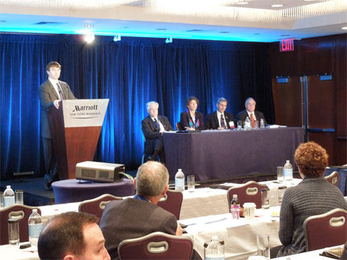
The Ultrasound First conference went on with tweets happening every hour! I was unable to attend this one, but so happy that my twitter friends did. It is obvious that 2013 truly is the year of ultrasound. Spreading to medical education, being a multi-disciplinary educational and practical tool, and having a united voice on its value were all discussed – in addition to some pearls on the hot topics including pelvic ultrasound and MSK ultrasound, as well as how ultrasound is becoming an acceptable tool for renal colic and breast masses.

Since I learn from all my Twitter friends, I figured the best way to share is to take out the middle person (yes, Im talking about me). That way you could get it from their own words: Here are only a few posts from #ACEP12  and #US1st that made me go “Hmmmm….” – with a little commentary every now again from me, because I just can’t NOT give my opinion – I know that’s shocking to those of you who know me. Heehee. My tweets are in here as well.

ACEP:

From @USEDCDN : Emergency US management course  “From Blaivas: Starting to see 1st lawsuits for lack of US use in vascular access” – This definitely sparked my attention – lawsuits for LACK of US use?? Wow, well the standard of care is changing, and if a proven tool to minimize complications is right next to you and you dont use it and that complication occurs… there’s a legal risk. Learn it, use it, love it and maximize patient safety.

Also from  “@USEDCDN: EUS MC Resnick: Emergency US is not an extension of physical exam. Big difference. It answers clinical questions.” Ok, this needs mention, but I already posted a rant about this – of course – so will not bother you with another rant… not right now, at least.

From @jeremyfaust  – “Weingart: 4. When is CPR futile? End tidal < 10 after 10 min. Confirm with US #acep12.” Enough said – and honestly, anything that Scott Weingart says, i will believe. period.

“Rice #ACEP12 echo in cardiac arrest- can see if cause PE/tamponade, or if standstill or beating heart. Look or you may waste time/resources” and “echo and IVC in critical patients: LV fxn, IVC collapse, RV size, contractility- will differentiate PE, CHF, hypovolemia, hypervolemia” – you never know what you may find, and what may be an intervention that you didnt think of until you saw your ultrasound (ie. tpa in a dilated RV).

@bedsidesono: lung #ultrasound talk from ACEP athttps://vimeo.com/51212231  brush up on A-Lines, B-Lines, lung sliding and more…#FOAMed” – what a giver he is! Stone is one to listen to, hear his opinion, and read his immense amount of publications.

Point of care US dominating new speakers forum so far at #ACEP12! Msk, soft tiss, pleural…”

Congrats @GeriaSonoMD on being new Chair of #ACEP12 US section mtg. Our fellow, Viveta Lobo said you talked her into EM. Awesome. So thx!

Raj Geria – new #ACEP12 US section Chair! Highest priority: pathway to US fellowship accreditation : to ensure safety and quality

Find @SAEMAEUS on twitter and follow to see what’s new with the ACademy

Nova panebianco at #ACEP12 taking about SAEM Academy of Emerg US and SonoGames – also subcommittees they are involved in..

A great resource from the new academy of emerg US : http://SAEM.org/academy-emergency-ultrasound-resources …

Resa Lewis #ACEP12 US section mtg- talking ACGME US milestones and how ACEP can help in achieving them for all residencies

Blaivas #ACEP12 – TEE will show potential causes of hypotension & shock ..Valvular dz..And can be electrically linked :pace & defibrillate. No need to interrupt chest compressions for TTE as can see what’s happening with heart from across rm c TEE. Can use TEE when bad view on TTE or unable to do TTE due to habitus, lung dz, chest compressions. TEE can assess quality of chest compressions too. TTE can tell you if there is standstill, clot in RA, dilated RV, tamponade, and to see a beating heart. TTE (echo) better than checking for pulses for need for chest compressions. AAMC mtg in SF! Spreading to med schools!. WINFOCUS and AIUM EM and crit care goals… Going global!

http://Sonocloud.org  and http://sonoguide.com  : 2 great online resources for images and education! – plus the test: http://emsono.com
@sinaiemus: Rob Blankenship at Ultrasound section meeting: over 56,000 ACEP US tests completed at http://www.emsono.com/acep/ACEP_EUS_Exam.html … #ACEP12
Congrats Vicki Noble and @ultrasoundpod for your well deserved award for your contribution to emerg ultrasound!!
Ultrasound First Forum:
  1. Jason T Nomura MD @Takeokun “To engage the patient groups you need people who are interested in patient advocacy not just the disease state. 
  2. View image on Twitter
  3.  Jason T Nomura MD @Takeokun “Lev demonstrating high res eval of ankle tendons with dynamic scanning for function, something that can’t be done with MRI 
  4. Jason T Nomura MD @Takeokun “Nazarian MRI does not have the resolution to evaluate the fibrillar pattern of the Achilles compared to US. 
  5. Jason T Nomura MD @Takeokun “Hoppmann- if education and integration of US starts in medical school it can change the paradigm. 
  6.  Jason T Nomura MD @Takeokun “Hoppmann has graduated several classes of medical students who had US integrated into their med school curriculum. 
  7. Jason T Nomura MD @Takeokun “Moreau most common imaging modalities for Team USA is X-ray and US, very little CT use.
  8. Mike Stone @bedsidesono “Levon Nazarian at  speaking on MSK imaging. It’s not just more convenient than MRI – higher res, no contraindications, pt’s prefer it”
  9.  Jason T Nomura MD @Takeokun “Nazarian US for sports med is portable to get the technology to the field and locker room, MRI not portable. 
  10. Jason T Nomura MD @Takeokun “Pellikka 2011 joint guidelines from ACC,ASE, ACCP and others about the appropriate times to use echo in the assessment of pts. 
  11.  Jason T Nomura MD @Takeokun “Moore bringing up the ASE and CV Anes guidelines for US guided vasc access. Advocates real time US guidance 
  12. Mike Stone @bedsidesono “Leslie Scoutt from Yale – ACR appropriateness criteria for recurrent renal colic – US & Noncon CT equal ratings 
  13.  Jason T Nomura MD @Takeokun “Scoutt 50% of pts with renal colic will likely have another episode.  that rad exp can build up.”
  14.  Jason T Nomura MD @Takeokun “Scoutt noncon CT is the “gold standard” for renal colic imaging in the US currently.  but there is the rad “risk.
  15. Joshua Copel @jacopel “Lynn Fordham (Pedi Rads) US optimal for pyloric stenosis now. No more need for upper GI or other radiation. 
  16. Jason T Nomura MD @Takeokun “Fordham N/V can be pyloric stenosis, malro, intussusception, and gastroenteritis. US for dx.
  17.  Jason T Nomura MD @Takeokun “IOTA group from Europe with close to 2,000 pts showed very good discrimination of malignant vs benign ovarian mass on US. 
  18. Jason T Nomura MD @Takeokun “IUD placement or misplacement easy to note on US; can present for DUB and pain. t
  19. Jason T Nomura MD @Takeokun “Advances to 3D US allows volumetric imaging that could only be done previously with CT or MRI. But US spares the radiation of CT 
  20. Joshua Copel @jacopel “ Dr. Beryl Benacerraf making case for US over CT, MR in female pelvic imaging at forum. pic.twitter.com/vCQYvpi8 View image on Twitter
  21. Jason T Nomura MD @Takeokun “ is not only about when &where US can be used but education for practitioners and patients per @AIUMPresAlfred
  22. Jason T Nomura MD @Takeokun “@AIUM_Ultrasound represents 9,200 members from 36 specialties with a focus on advancing US use 
  23.  Joshua Copel @jacopel “ opening of US First forum now at Marriott NYC. Over 100 attending from medical profs, industry, payors. Very exciting & energetic”
  24.  Jason T Nomura MD @TakeokunIt does seem to be a who’s who of US at the reception.

SonoPearls… from Stanford Bedside Ultrasound Course… for FAST, Echo and RUSH

Teaching US with fellow US lovers is just too fun. What I appreciated most – multi-specialty!! …

…with Sarah Williams (EM), me (EM), Zoe Howard (EM), Brie Zaia (EM), Darrel Sutijono (EM), Phil Perera (EM/IM), Yoshi Mitarai (EM-ICU), MyPhuong Mitarai (EM), Viveta Lobo (EM), Anne-Sophie Beraud (Cards), John Kugler (IM):

Multi-dept instructors: EM, IntMed, ICU, Cards.
Multi-dept learners: Anesth, IntMed, Peds, Surg, EM, NPs

US Pearls: Continue reading

ULTRAFEST in Southern CA for medical students!

ultrafest.org – have to give props to the creator of my US itch, Dr. Chris Fox at UCI (my prior stomping grounds for EM residency) for his hugely successful med stud US course – over 200 students!! May 20, 2012 – Chris, Im looking forward to getting some pics from that! Wish I could have been there!