SonoApp- Bedside ultrasound for thyroid nodules? – a patient’s question and experience

This post involves a personal experience from a free lance writer who asked me whether we (EM or primary care docs) should/can be doing bedside ultrasound for an assessment of the thyroid when we feel something wrong – even if its for the sole purpose of expediting care and biopsy, or alleviating concerns with a negative study.  It was a valid question, and she wrote about her experience below. The thyroid is not a difficult gland to find, nor is it a technically difficult ultrasound to perform – although not a current use for bedside ultrasound, it may start to be given the ease to which nodules can be seen: (for pictures of normal thyroid and abnormal thyroid nodules, see the end of the post  – the pics are not her thyroid)

Point-of-Care Ultrasounds: Can They be Used for Thyroid Nodules?

At a regular physical exam, my doctor discovered a small lump in my neck. She said it was on my thyroid, and referred me to an endocrinologist for further evaluation. At only 19 years old, I was scared and extremely uneducated in regards to thyroid health and diagnoses. My endocrinologist ordered a radiology ultrasound to be done at a later time where I had the technician hovering over me. After the doctor declared I had two nodules, I was scheduled to go back yet again for an ultrasound-guided fine needle aspiration (FNA) to help rule out cancer. This persistent waiting from one appointment to another for small amounts of information were frustrating, and I wondered why my own doctor couldn’t just do the initial ultrasound to visualize the nodules, streamlining the process and expediting my work up.

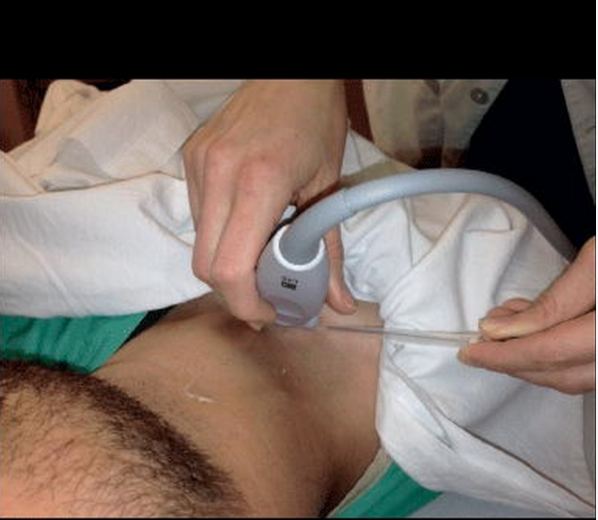
The day finally arrived for the FNA, and the scene couldn’t be more uncomfortable—and comical at the same time. As the technician tried to gain an image of the right spots with the large wand, the endocrinologist struggled to bypass the size of it in order to gain the samples he needed. On top of that, it was nearly four months from the time of my original physical exam before I found out I had benign nodules. I was told I would need to follow up with a radiology ultrasound every few years.

Point-of-care (POC) ultrasounds, units that are small and possibly hand-held that are increasing in popularity among physicians and emergency doctors, can used by primary care doctors with their assessment. Not only are they portable, but they are easier and more comfortable to use for all parties involved. While not widely publicized or used for thyroid nodules yet, there is potential that POC ultrasounds could aid in diagnosis and follow-up for related patients.

Defining Thyroid Nodules

Thyroid nodules are areas of abnormal cell growths within the thyroid gland. Depending on their size, nodules may form a visible mass in the neck. Some can also cause a goiter, or enlarged thyroid gland. The majority of thyroid nodules are not cancerous. However, an ultrasound and FNA can rule this out. These nodules may go undetected for years. Others can coexist with other related issues, such as hypothyroidism and hyperthyroidism. Thyroid nodules can also become painful to the touch and when you swallow.

Unless you can feel a lump in your thyroid gland, these types of nodules are first detected through a physical exam of the neck. Conventional radiology ultrasounds are currently used as a follow-up to locate the nodules and rule out any potential problems.

Role of POC Ultrasounds

On a larger scale, the increased use of POC ultrasounds can help the health of patients. It can even help save lives: although rare, thyroid cancer can be deadly if not caught early. Most cases of thyroid nodules and masses occur in older patients, some of whom may not be able to make it to radiology offices for a traditional ultrasound. By employing the use of a POC ultrasound, a physician can quicken the diagnosis and make the process more comfortable for patients. The fact is that once patients leave their primary care physicians’ offices, it is up to them to follow up on referrals to ultrasound appointments. Having a POC device handy ensures that a patient won’t miss these crucial diagnoses.

Having an ultrasound for thyroid nodules is easier with POC ultrasounds. Since the area of investigation is small, this approach makes the most sense when considering comfort for both doctor and patient.

Another benefit is the cost. POC ultrasounds cost providers less money, and it may be more affordable to patients. With the rising costs of healthcare, more and more patients are missing out on critical exams. By making thyroid scans more affordable, patients may be less likely to skip out on their appointments. The downside is that POC ultrasounds are not used for thyroid nodules yet. By pushing for their use in all areas of healthcare, POC may ease the ultrasound experience and even save lives.

Resources

Kristeen Cherney

Author Bio: Kristeen Cherney is a freelance health and lifestyle writer who also has a certificate in nutrition. Her work has been published on numerous health-related websites.

Pics from aium.org – NORMAL ADULT THYROID

normal thyroid

Pics form ultrasound-images.com :

ADENOMAS

thyroid_adenomatous nodules thyroid_follicular adenoma

FOLLICULAR ADENOMAthyroid_follicular adenomas

PAPILLARY CARCINOMAthyroid_papillary carcinoma


SonoCase: 46yo shortness of breath – guest post by Jacob Avila @UltrasoundMD #FOAMed

Our newest guest post is by one of the best emergency medicine resident educators I know – of course, you dont want to miss his educational pearls on twitter too – Dr. Jacob Avila. He discusses a case that illustrates how bedside ultrasound can help in your unexplained short of breath patient, and even cancel that triage bias that your attending can do to sway you away from the truth. Let’s give it to Dr. Avila for highlighting (with a great literature review) how ultrasound can help you too. Here it is – enjoy! (note: not all images were of this patient, but were taken from other resources)

“You arrive to the emergency department for your first night shift of the month, and as you place your bag on the desk, the attending walks towards you with a chart in his hand. “Do you mind seeing this patient? It’s a COPD’er with dyspnea.  It’s probably just a COPD exacerbation.”  You look at the chart and see that it’s a 46 year old female with shortness of breath.  As you walk into the room, you notice the patient appears slightly pale, is afebrile, has an O2 saturation of 91% and is tachycardic in the 110’s with a blood pressure of 105/76, temperature of 98.5° and respiratory rate of 26.  While taking the history, you note that the patient is a smoker and recently returned from a 12 hour car ride to see relatives.  Suspecting that this may be something other than simple COPD exacerbation, you grab your ultrasound machine and start with the cardiac echo (as described in the RADiUS protocol) and are able to get the following image:

This apical 4-chamber view shows severe right heart dilation, defined as a RV:LV ratio >1.  However, you remember that the patient has a history of COPD, and chronic pulmonary hypertension can cause chronic right ventricular dilation1.  At that moment, the patient becomes hypotensive with a systolic blood pressure in the 70’s and develops severe respiratory distress. What should we do?

Early diagnosis of a pulmonary embolism (PE) is exceedingly important, as two thirds of patients with mortality associated with a PE die within the first hour of their presentation2, and intuitively, those who are treated earlier generally have a better prognosis 3.  The definitive diagnosis of a PE requires the use of a CT scanner 4, but in a patient who is unstable, like this one, that isn’t an option. Looking at the right ventricular to left ventricular ratio is a maneuver that can rapidly change your differential diagnosis or confirm what you previously suspected. A recent study by Dresden et al found right ventricular dilatation identified by emergency physicians had a specificity of 98% for a PE.  That number is impressive, but when you look at the methodology section of the publication, only 10% of the patients they included had coexisting COPD, and all of the false positives in the study were in patients with COPD5.   One  technique that may help differentiate between chronic and acute dilation is looking at the RV free wall in the subxiphoid view while in end diastole. A free wall size >0.5 cm is more likely to be chronic RV dilation6i. However, this view is not always possible in all patients.  Another echo sign you could look for is the McConnell sign (apical winking of the right ventricle during systole), which previously was reported to have an impressive 94% specificity and 77% sensitivity for an acute PE7l, but a subsequent and larger study found the McConnell sign to be only 70% sensitive and 33% specific for a PE8. Take a look at what the McConnell sign looks like”


Another, less commonly seen finding would be directly seeing the clot in the right atrium (RA) or in the pulmonary arteries.

Clot in RA:

RA clot labeled

Clot in pulmonary artery:

Pulm clot labeled

Of more practical use are two other sonographic findings: Deep venous thrombosis (DVT) and distal pulmonary infarction.

In a study that included 199 examinations, bedside 2-point compression evaluation of the greater saphenous/femoral vein junction and the popliteal veins of patients with suspected DVT was found to be 100% sensitive and 99% specific for DVT 9.   However, it is possible for a patient to present with an acute PE and have a negative DVT, and only about 40-50% of patients with DVT’s will end up having a PE10, 11

DVT on one side diagnosed by noncompressible vein:

More recently, lung ultrasound has been explored for the assessment of a suspected PE. A recent systematic review and meta-analysis by Squizzato et al which included 10 studies and a total of 887 patients found lung ultrasound to have a mean sensitivity of 87% and a mean specificity of 82% for acute PE12.  What they looked for in the lung was the presence of triangular, wedge or rounded hypoechoic, pleural based lesions.  These lesions are thought to be due to embolic occlusions that resulted in either focal atelectasis with extravasation of blood or focal infarction of the lung parenchyma . However, they state in their publication that “Several methodological drawbacks of the primary studies limit any definite conclusion”.

Lung infarction:


Instead of looking at just one specific sonographic finding for the diagnosis of acute PE, a better method may the use of multi-organ sonography.  Recently, Nazerian et al. published a study utilizing multi-organ sonography in the diagnosis of PE.  This study used echo, lung and DVT ultrasound to diagnose PE and found that when the three ultrasounds were combined, they yielded a sensitivity of 90%, which was significantly higher than each of the exams by themselves13.

Like any physical exam finding, lab reports or other radiographic assessments, the sonographic analysis of a patient with a suspected pulmonary embolism should be used as part of your diagnostic quiver, and not the silver bullet.  Any of the above mentioned ultrasound findings of acute PE can potentially be found in other,  non-PE causes of dyspnea.  DVT’s can just be DVT’s, RV enlargement can be chronic or from an RV infarction, and subpleural fluid collections can be seen in contusions, pneumonia  and cancer.  This doesn’t mean not to use it though.  Just think about all the other tests we use in the emergency department, such as EKG’s, chest x-rays, troponins, BNP, and the d-dimer.  All of these can be abnormal in PE and in non-PE entities.

Now back to our patient.  She is a 46 year-old female with COPD that had right heart enlargement, which we learned above can be  seen in COPD without the presence of a PE.  You were unable to get a good subcostal view of the heart to measure the lateral wall, mostly because the  patient did not tolerate being laid flat.  You move on to the lungs and in the lower right thorax and there you find two hypoechoic, pleural based lesions.  Heparin and a CT scan are ordered, and the CT scan shows a large clot located in the right main pulmonary artery.

Here is the CT scan showing the clot:

Avila_Clot in pulmonary artery CT

To see a recent podcast by Ultrasoundpodcast on multi-organ US for PE, go here.

References:

  1. Otto, Catherine M.. Textbook of clinical echocardiography. 5th ed. Philadelphia, PA: Elsevier/Saunders, 2013. Print. p 247
  2. Wood KE. Major pulmonary embolism: review of a pathophysiologic approach to the golden hour of hemodynamically significant pulmonary embolism. Chest. 2002;121:877-905
  3. Jelinek GA, Ingarfield SL, Mountain D, et al. Emergency department diagnosis of pulmonary embolism is associated with significantly reduced mortality: a linked data population study. Emerg Med Australas. 2009;21:269-276
  4. Goldhaber SZ, Bounamenaux H. Pulmonary embolism and deep vein thrombosis. Lancet 2012:379:1835-46
  5. Dresden S1, Mitchell P2, Rahimi L2, Leo M2, Rubin-Smith J2, Bibi S2, White L3, Langlois B2, Sullivan A4, Carmody K5 Right ventricular dilatation on bedside echocardiography performed by emergency physicians aids in the diagnosis of pulmonary embolism. Ann Emerg Med. 2014 Jan;63(1):16-24. doi: 10.1016/j.annemergmed.2013.08.016. Epub 2013 Sep 27.
  6. Rudski LG, Lai WW, Afilalo J, Hua L, Handschumacher MD, Chandrasekaran K et al. Guidelines for the echocardiographic assessment of the right heart in adults: a report from the American Society of Echocardiography endorsed by the European Association of Echocardiography, a registered branch of the European Society of Cardiology, and the Canadian Society of Echocardiography. J Am Soc Echocardiogr. 2010;23:685-713
  7. McConnell MV, Solomon SD, Rayan ME, Come PC, Goldhaber SZ, Lee RT. Regional right ventricular dysfunction detected by echocardiography in acute pulmonary embolism. Am J Cardiol 1996;78:469e73.
  8. Casazza F, Bongarzoni A, Capozi A, Agostoni O. Regional right ventricular dysfunction in acute pulmonary embolism and right ventricular infarction. Eur J Echocardiogr. 2005;6:11-4
  9. Crisp JG, Lovato LM, Jang T. Compression ultrasonography of the lower extremity with portable vascular ultrasonography can accurately detect deep venous thrombosis in the emergency department. Ann Emerg Med. 2010;56:601-610
  10. Kearon C. Natural history of venous thromboembolism. Circulation. 2003;107:22-30
  11. Moser KM, Fedullo PF, Littlejohn JK, Crawford R. Frequent asymptomatic pulmonary embolism in patients with deep venous thrombosis. JAMA 1994;271:223-225
  12. Squizzato A1, Rancan E, Dentali F, Bonzini M, Guasti L, Steidl L, Mathis G, Ageno W. Diagnostic accuracy of lung ultrasound for pulmonary embolism: a systematic review and meta-analysis. J Thromb Haemost. 2013 Jul;11(7):1269-78. doi: 10.1111/jth.12232.
  13. Nazerian P, et al. Accuracy of Point-of-Care Multiorgan Ultrasonography for the Diagnosis of Pulmonary Embolism.Chest. 2014 May 1;145(5):950-7. doi: 10.1378/chest.13-1083

SonoCase: 46yo c/o abdominal pain h/o cocaine use – sup mesenteric art dissection #FOAMed

Drs. Davis and Kendall write up a very interesting case in the Aug 2013 issue of Journal of EM where the ultrasound made the diagnosis, quite easily too. They discuss a 46 year old male with a history of current cocaine use AND a prior history of an aortic dissection, of course, who was complaining of sudden onset of abdominal pain and found to be severely hypertensive. The diagnosis on the top of their list was aortic dissection/aneurysm/rupture – and when they looked, they saw even more. Below is the abstract:

Background

A timely diagnosis of aortic dissection is associated with lower mortality. The use of emergent bedside ultrasound has been described to diagnose aortic dissection. However, there is limited literature regarding the use of bedside ultrasound to identify superior mesenteric artery dissection, a known high-risk feature of aortic dissection.

Objective

Our aim was to present a case of superior mesenteric artery dissection identified by bedside ultrasound and review the utility of bedside ultrasound in the diagnosis of aortic emergencies.

Case Report

We report a case of superior mesenteric artery dissection found on emergent bedside ultrasound in a 46-year-old male complaining of abdominal pain with a history of cocaine abuse and prior aortic dissection. Bedside ultrasound in the emergency department revealed an intimal flap in the descending aorta with extension into the superior mesenteric artery prompting early surgical consultation before computed tomography because of concern for acute mesenteric ischemia.

Conclusion

Superior mesenteric artery dissection is a high-risk feature of aortic dissection and can be identified with emergent bedside ultrasound.

Just one of their images is displayed below – but take a look at the video in JEM to truly see the awesomeness. A subscription and password is required, but it’s a great journal with lots of cool ultrasound cases published almost every month.

Screen Shot 2013-09-03 at 9.54.59 AM

SonoCase: 60yo blunt chest trauma, 82yo with chest pain- in JEM by @ultrasoundREL

In a recent article in the Journal of Emergency Medicine, Dr. Resa Lewiss and friends, discuss 2 cases of thoracic aortic aneurysm identification by focused cardiac ultrasound. It is a great case report that highlights the need to include the aortic root and descending thoracic aorta in the parasternal long view of your focused cardiac echo.

“A 60-year-old man presented to the emergency department (ED) after a blunt traumatic injury to his back while at work. During the focused cardiac ultrasound examination, the aortic outflow tract distal to the aortic valve appeared enlarged and the aortic root measured 5.49 cm.

Screen Shot 2014-03-04 at 9.16.55 AM

“An 82-year-old man with hypertension presented to the ED with 1 month of chest pain radiating to the back. The focused cardiac ultrasound examination demonstrated enlargement of the descending thoracic aorta at 4.82 cm.”

Screen Shot 2014-03-04 at 9.17.06 AM

SonoCase: 60yo in cardiac arrest in @EPMonthly by @TeresaWuMD @TheSafetyDoc #FOAMed

In the recent issue of Emergency Physician’s Monthly (one of my favorite EM magazines), Drs. Teresa Wu and Brady Pregerson once again hit the ultrasound wave and start soaring in their newest insert describing the utility of bedside ultrasound during cardiac arrest and post-mortem.

They describe it best: ” ….60-year-old male who collapsed at work and remained unresponsive. They state that there was bystander CPR and a lot of freaking out by coworkers. The only past history they have was from a coworker who thought he had high blood pressure. There was also a witness who told them he was just walking, then doubled over and collapsed without saying a thing. No one knew if he had any symptoms earlier in the day. Paramedics state he was initially in a PEA rhythm at a rate of 120 bpm on the monitor. They started an IV, gave him a 500cc saline bolus, intubated him, and have given three rounds of epi. They estimate a 15 minute down time prior to their arrival and a 10 minute transport time with no return of spontaneous circulation. In fact, things are going in the opposite direction as he has been in asystole for the past five minutes.

They move him onto the bed where your EMT takes over CPR. You note good and symmetric assisted breath sounds via the ET tube, but minimal palpable femoral pulse despite what appears to be good CPR to the tempo of the Bee Gees hit “Staying Alive”. On the monitor there is asystole in two leads. Pupils are fixed and dilated despite no atropine having been received. Things are not looking promising.

You request saline wide open and a final round of epinephrine while you take a look for cardiac motion with the ultrasound machine. To minimize interruption of CPR you don’t have the EMT pause until you are completely ready to look. You also have the RT hold respirations to avoid any artifact. There is no cardiac motion. You verbalize this to your team. The heart does not appear dilated and there is no pericardial effusion. You ask aloud, “anyone have any other suggestions” prior to calling the time of death.

Of course you next wonder what did him in: MI, PE, something else… His belly looks pretty protuberant, so you decide to take a quick look at his abdomen to check for free fluid. What you see is shown in the two images below. ”

Screen Shot 2013-08-27 at 6.26.16 PM

What do you think killed this gentleman? Trust me, you want to read more and see what exactly the ultrasound image is  – as it is quite an interesting finding: go here.

For a discussion from a prior post on ultrasound during cardiac arrest, go here.

SonoCase and Procedure: Supraclavicular Subclavian lines from @EMNews #FOAMed

In a recent issue of EM News where Dr. Christine Butts once again offers insightful advice on how bedside ultrasound can assist with patient care, she gets help by one of her residents from Louisiana State University, Dr. Talbot Bowen. They offer their insights into ultrasound guided SUPRAclavicular subclavian lines – yup, that’s right – read below as they say it best:

“Ultrasound guidance in supraclavicular subclavian vein (SCV) catheterization is a relatively new concept. Traditional infraclavicular SCV catheterization is poorly amenable to ultrasound guidance because of the overlying clavicle, which can make visualization and direct guidance difficult. Supraclavicular SCV catheterization for central line placement has several advantages: practicality in cardiopulmonary arrest, decreased incidence of central line infections, lower risk of pneumothorax, and decreased incidence of thrombosis……..Once the subclavian vein is identified, the central venous catheter may be placed by dynamic ultrasound guidance. The introducer needle is advanced with gentle negative pressure from the end of the transducer. (Image 3.) This “in-plane” approach allows the operator to visualize the needle and needle tip at all times while advancing toward the vessel.”

Screen Shot 2013-08-27 at 5.40.56 PM

Read the entire entry to get even more of the goodness they describe, here.

For a post on all ultrasound guided procedures, go here.

SonoCase: 25yo unresponsive, found down – by @KasiaHamptonMD #FOAMed #FOAMus

In case you all were unaware, Dr. Kasia Hampton is REALLY into ultrasound. She is a resident in emergency medicine and is teaching her colleagues how to use it. She has case after case of great findings, quick pick-ups, and lives saved and management changed due to that little old ultrasound machine. She even has another twitter/blog, called @tres_EUS  – a site for residents interested in ultrasound cases/leadership/research/etc. She emailed me this case that I thought was a fabulous use of ultrasound and actually shows what I harped on and on about with EMCrit on a recent podcast on FAST scans highlighted in our SonoTips and Tricks on FAST scan upper quadrants.

Enjoy!

“25 yo male was found unresponsive per bystanders. Upon EMS arrival he was noted to have multiple stab wounds to the upper extremities and chest. Initial set of vitals revealed tachycardia without hypotension. Patient was intubated at the scene “for airway protection”. Mechanically ventilated upon ED arrival with the following vitals: BP 135/90 mmHg, HR 105 BPM, respirations 16/min, SpO2 100%, T 35.8 C. GCS 3T. During secondary survey found to have one stab wound to the left anterior chest (inferior to the nipple), and second stab wound to the right posterior chest (lateral to the inferior aspect of the scapula). Additional two stab wounds to both shoulders were superficial and were no longer bleeding. No apparent abdominal (wall) injuries were noted. Abdomen was non-distended and soft.

The RUQ FAST scan:

Seek and ye shall find 3

FAST ultrasound evaluation was performed after the patient was log-rolled in both directions – first to the left and then to the right.  Subsequently the patient was taken to CT scan. He remained hemodynamically stable. Below the comparative findings of FAST vs CT scans.

IMAGING

FAST ULTRASOUND

CT

RUQ

perihepatic free fluid

perihepatic free fluid

SUBXIPHOID

no pericardial effusion

no pericardial effusion

LUQ

no free fluid

trace perisplenic free fluid

PELVIC

no free fluid

no free fluid

Given stab wound to left anterior chest with presence of free fluid in the abdomen (with hepatic and splenic injuries identified on CT), patient was taken to the operating room. Injury to pericardium itself without pericardial effusion was suspected on CT. During the surgical exploration it appeared that the stab wound to the left chest only nicked the pericardium (no blood within pericardial sac), while penetrating the left diaphragm, left lobe of the liver, stomach, spleen and pancreatic body.

This case illustrates a few important concepts:

  1. The ultimate importance of visualizing the paracolic gutter around inferior pole of the right kidney on FAST ultrasound exam;
  2. The dilemma of performing FAST scans after the patient has been log-rolled (in particular to the left side, while less important if rolled onto the right);
  3. The superiority of Secondary UltraSonographic Survey In Trauma (SUSS IT) over clinical exam for non-suspected injuries.

4 @broomedocs with love - SUSS IT OUT

In this particular case I wonder if the trace perisplenic free fluid would have been identified on FAST performed before log-rolling? Additionally, it is quite amazing how misleading was the clinical secondary survey in comparison to FAST findings and intra-operative discoveries. “

SonoCase: 72yo back pain & hypotensive – by Dr. Calvin Hwang @helixcardinal #FOAMus #FOAMed

Another great guest post! – by Dr. Calvin Hwang, aka @helixcardinal  – as well as the senior resident at Stanford/Kaiser EM program who updates the @StanfordEMRes residency twitter feed, provided an excellent case that illustrates a reason/indication to perform bedside ultrasound – especially the Echo/IVC and Aorta applications – illustrating why these applications are imperative to the RUSH protocol – along with good clinical judgement. Enjoy!

“Code 3 ringdown from EMS: 70 yo F coming in with 3 days of chest, back and abdominal pain, hypotensive with SBP in the 70s.

On arrival, patient is grimacing in pain, pale, diaphoretic.  She is otherwise healthy with no past medical history.  Just arrived from Thailand 1 week ago to visit her daughter and had been complaining of pain in her chest, back and abdomen.  Went to a primary care physician where she was noted to be hypotensive and sent to the ED.

Initial vital signs: BP 73/30, HR 110, T37.0, RR 25

Screen Shot 2013-08-22 at 6.21.37 PM

With the trusty bedside ultrasound, I immediately went to where I thought would be the diagnosis: ruptured AAA…..but…..

The abdominal aorta scan : I was shocked when I noticed it to be of normal caliber.  Nevertheless, I worked my way up the abdomen to the subxiphoid view when I saw:

Though it was atypical for the patient to be hypotensive and tachycardic, the presence of a pericardial effusion without tamponade suggested aortic dissection to me.  My attending got on the phone to prepare to transfer the patient while I contacted the radiologist to clear the CT scanner.  Though I attempted to view the descending aorta and aortic outflow tract on a more focused echo in the brief interim through a parasternal approach, I was unable to obtain good windows.  The IVC was plump and the rest of the FAST was negative.  A quick Chest XR was done:

Screen Shot 2013-08-22 at 6.21.01 PM

…..which did not show a wide mediastinum according to radiology.  The patient was whisked away to the CT scanner and within 45 minutes of ED arrival, the diagnosis of a Stanford type A aortic dissection with pericardial effusion (but not tamponade) was confirmed.  This would not have been possible without bedside ultrasound as I think most clinicians would have been falsely reassured by the normal CXR (widened mediastinum only present in 60% of aortic dissections1).

The patient was fluid resuscitated with crystalloid, her BP improved to 100/60 and HR came down to the 80s.  While awaiting transport, I attempted to place an arterial line for close BP monitoring.  However, approximately 60 minutes after ED arrival, the patient became progressively bradycardic and coded.  My institution’s cardiothoracic surgeons were already at bedside and performed a sternotomy with pericardial window.  Despite our efforts, we were never able to obtain return of spontaneous circulation and the patient was pronounced. These patients rarely make it to the ED due to how quickly they can decompensate, but if they do, quickening the diagnosis may help get them the intervention they need (clinical suspicion and appropriate use of bedside ultrasound is key), although a high mortality still exists.

  1. Aldeen A, Rosiere L.  “Focus on: Acute Aortic Dissection.” ACEP News, July 2009.

SonoCase: 32yo shortness of breath – by @Medialapproach #FOAMed

We have had some great additions of guest posts of cases where ultrasound mattered and helped with their diagnosis and treatment. Below is a case from Vince DiGiulio, an EMT and ED tech extraordinaire and more! – also known as@MedialApproach of the medialapproach.com as well as the founder of a great Google+ account on ultrasound. Read his case below and enjoy!
“In this case I was able to nail down the cause of the patient’s symptoms in 5 minutes, and I’m only an EMT whose US teaching has come entirely from online resources like SonoSpot. Here’s the story:

It’s a hot summer’s day and you are working a busy shift in the Minor Care unit of a community ED when a 31 year-old man presents with a chief-complaint of shortness of breath (SOB).

He states that he has been feeling SOB on exertion for the past 3-4 weeks, having attended the walk-in two weeks prior with the same complaint. There he was diagnosed with asthma and given an albuterol MDI, a course of PO steroids, and also a course of PO azithromycin “in case it was something more.” His symptoms had not improved so he decided to attend the ED for another opinion.

From the doorway you see a moderately overweight (5’9” 200#) Caucasian male in no acute distress. He is exhibiting a normal respiratory rate with no elevated work of breathing. His skin is warm and of normal color, but upon closer inspection you’re a bit surprised to notice he is actually moderately diaphoretic. He chalks it up to the outside temperature of 90 F, but it’s a chilly 70 F in the department and he’s been seated in bed for at least 20 minutes. “Hmmm,” you say to yourself.

Vital signs at rest are as follows: HR 115 bpm, RR 20/min, BP 122/68 mmHg, Temp 37.1 C.

On auscultation he has a bit of bi-basilar rales.

After obtaining a history, you head back to your desk to enter some orders when you see that an ECG and CXR were already performed at triage.

32yo M - SOB on Exertion x 3 wks_ECG

[http://sonocloud.org/files/photos/1373606099f1a0ab_o.jpg]

The ECG shows sinus tachycardia, left-atrial abnormality, left-axis deviation, poor R-wave progression, large S-wave in the right-precordial leads, and secondary ST and T-wave changes. This picture is consistent with left-ventricular hypertrophy.

Screen Shot 2013-12-02 at 12.58.36 PM

http://sonocloud.org/files/photos/13736928941892d4_o.jpg]
The CXR was read by radiology as “mild-to-moderate cardiomegaly, new from prior film (2 years ago), consider pericardial effusion.”

This Minor Care case is starting to get a bit more complicated and you’re beginning to wish you had seen the patient with chronic low-back pain instead. Not quite sure what to make of this patient’s presentation and afraid of backing up the whole department while you try to make a hard-sell on this young, otherwise healthy patient to cardiology, you instead grab your trusty ultrasound machine and head for the bedside. Here is what you see.

[http://sonocloud.org/watch_video.php?v=NAHA61SSH3B7]

In this apical 4-chamber view, you first notice that all four chambers are markedly dilated and hypokinetic. Looking specifically at the left-ventricle, it exhibits with severe hypokinesis throughout, maybe with a touch of apical akinesis. Looking closely at the apex of the LV also shows that there is an apical mural thrombus, often seen in patients with akinesis or severe hypokinesis of that region.

 [http://sonocloud.org/watch_video.php?v=SG794W7MBYBG]

As evidenced by the obvious blue jet in the left-atrium, this color-Doppler image of the mitral valve demonstrates significant mitral regurgitation. At formal echocardiography it was graded as “moderate, 2+ mitral regurgitation.”

32yo M Mitral Doppler

[http://sonocloud.org/files/photos/1373605496135c3c_o.jpg]

This pulsed-wave Doppler image shows monophasic flow through the mitral valve with a nearly absent A-wave. This proves that in addition to systolic dysfunction, the patient has significant diastolic dysfunction as well in a restrictive pattern.

[http://sonocloud.org/watch_video.php?v=SK85U7KAMSW1]

Here is a mid-ventricle parasternal short-axis view that further demonstrates the global hypokinesis of the left ventricle. It is also clear that the ventricle is large and dilated, but not hypertrophied. In this patient’s Cardiology echo, his ejection fraction was estimated in the range of 10-15%. I’m an inexperienced echocardiographer, but in addition to global hypokinesis I might specifically see some anterior-wall akinesis here as well.

[http://sonocloud.org/watch_video.php?v=KGUYWXGR2YG2]

This parasternal long axis view offers a final example of the patient’s global hypokinesis, along with a nice shot of the mitral valve. The aortic valve is also in view, but not clearly seen. Notably there is also no sign of pericardial effusion, often visible in this view if present.

So what’s our final impression? Summarizing all of the specific findings listed above, this patient has a dilated cardiomyopathy. While the workup and management of this patient could encompass a week’s worth of posts, here are the main take-home points from this case:

  1. Beware patients who are diaphoretic or tachycardic at rest. Afebrile and in no acute distress, it became essential to find a source of this patient’s few abnormalities on physical exam.
  2. Don’t be afraid of ultrasound in the Minor Care department. We like to talk a lot about the utility of ultrasound during a patient’s resuscitation, but it can be equally useful in an ambulatory setting as well.
  3. Bedside ultrasound expedites care. Without bedside ultrasound this patient would have been waiting around hours (or days) for a formal echo, if it was going to be performed at all.
  4. Sell! Sell! Sell! In most circumstances cardiology would have been very reluctant to come see an otherwise healthy 31 year-old patient, but in this case the bedside images provided immediate and definitive proof that the patient needed specialty care. It also probably gained us some street-cred with the cardiologist who could look at the saved images right in the department.
  5. Shoot first, ask questions later. In a case like this, there is no need to perform an extensive interpretation of your images at the bedside. From the very first view it was clear the patient had a dilated cardiomyopathy, so cardiology was immediately paged and the patient was readied for admission. During that time additional views were quickly obtained for later evaluation, but that first shot told us all we needed to know to make a disposition on the patient.
    As an ECG nerd, I liken it to reading the tracing of a patient with a profound wide-complex tachycardia. At the bedside there is rarely any need to get too fancy differentiating VT from SVT with aberrancy since the WCT algorithm is safe and effective for both, but once the patient is stabilized I can then go back and look for signs of AV-dissociation on the ECG to really prove it was VT.
    You think I noticed the apical thrombus in this patient’s AP4 view? No-way! That’s something Mike Mallin of the Ultrasound Podcast picked up for me when I shared the case with him. I didn’t even know how to read a pulsed-wave Doppler at the time I met the patient, but I knew how to capture the image at the level of the mitral valve so that I could review and learn from it later.

Anyway, thanks to bedside US (and you!) this patient ended up having his dilated cardiomyopathy recognized and promptly treated. Without these surprising images there’s a really good chance this patient would have been symptomatically treated for his SOB in the ED and then discharged back home. If anything, being able to reference these clips gave our emergency physician a very strong card to play in getting cardiology to take the case seriously.”

SonoCase: Renal Ultrasound for Renal Colic: a cost/benefit analysis? by @EPMonthly #FOAMed

Once again, Drs. Teresa Wu and Brady Pregerson do an excellent job in highlighting a case in EP Monthly (and a topic that I am so incredibly passionate about – not only because of the benefit to the patient, the minimizing of CT scans/radiation, and the time spent in its work up – but also in health care cost and expediting diagnosis and management.) What am I talking about? Well, RENAL ULTRASOUND for RENAL COLIC. Yeah, I know, it sounds obvious. But, I heard of a patient the other day (again!) who had a known history of kidney stones, who had the same pain as her prior kidney stone flank pain, who begged to not have yet another CT scan done since it would have been her 13th for this at the age of 40. I highlighted this topic and other studies on it in a prior post, and AIUM posted a sound judgment series written by Drs. Chris Moore and Leslie Scoutt on this topic too.

So, let’s talk about TWu and Brady’s addition to the mix. Of course, they always start off their case with humor, yet reality, by saying : “I have to do a cost-benefit analysis of the situation,” your eager intern replies. It’s the end of the academic year and you are forcing your soon-to-be R2s to become more autonomous and confident in their management plans. You are amazed at the various answers you now get when you ask the simple question, “What do you want to do?” You ask your intern to summarize the case for you. He just finished evaluating a 21-year-old male who presented to your ED with back pain. The patient states that his “back is killing him” and he thinks he strained his muscles working out too hard at the gym last week. He just started doing CrossFit and he’s worried that he overdid it. The patient notes that the pain is 10/10 and that he has had minimal relief with his friend’s Vicodin. He’s tried icing his back and even sat in the hot tub all weekend per his friend’s recommendation. Nothing is working so his friend told him to come into the ED to get a prescription for something “stronger.”……

“Your question about whether or not this young 21 year old needs any imaging is giving him pause. “I think the cost of the imaging and the risk of radiation are too high. I don’t think there’s much benefit to keeping the patient here any longer. Plus I don’t know what we’d be looking for,” he replies. You are happy with your intern’s logic and pop into the room to see the patient. Within seconds, you realize that Vicodin and a hot tub probably won’t fix this patient’s pain. The patient is sitting hunched over on the stretcher rocking back and forth in pain. He has no appreciable tenderness to palpation over any of his back muscles, and there is no asymmetry or tightness on your exam. You are unable to reproduce or worsen his symptoms with testing his range of motion, but he is definitely rubbing his right lower back to try to ease his pain. You walk out of the patient’s room and grab your intern and the ultrasound machine. As you head back towards the patient’s room, you pimp your intern on the other more serious causes of low back pain. Acknowledging that you have the ultrasound machine in tow, your clever intern starts rattling off the diagnoses that can be easily made with bedside ultrasound. AAA, atypical appendicitis, cholecystitis, nephrolithiasis, abscess, etc. Since the patient is sitting upright and hunched over in pain, your intern decides to start his scan with a view of the right flank….”

Screen Shot 2013-07-17 at 2.37.30 PM

BAM! oh yeah – do you see it? Weren’t expecting that? Funny what happens when you look, right? You must read about their findings and the pearls and pitfalls of renal ultrasound – go here for the true meat of the article.