Our newest guest post is by one of the best emergency medicine resident educators I know – of course, you dont want to miss his educational pearls on twitter too – Dr. Jacob Avila. He discusses a case that illustrates how bedside ultrasound can help in your unexplained short of breath patient, and even cancel that triage bias that your attending can do to sway you away from the truth. Let’s give it to Dr. Avila for highlighting (with a great literature review) how ultrasound can help you too. Here it is – enjoy! (note: not all images were of this patient, but were taken from other resources)
“You arrive to the emergency department for your first night shift of the month, and as you place your bag on the desk, the attending walks towards you with a chart in his hand. “Do you mind seeing this patient? It’s a COPD’er with dyspnea. It’s probably just a COPD exacerbation.” You look at the chart and see that it’s a 46 year old female with shortness of breath. As you walk into the room, you notice the patient appears slightly pale, is afebrile, has an O2 saturation of 91% and is tachycardic in the 110’s with a blood pressure of 105/76, temperature of 98.5° and respiratory rate of 26. While taking the history, you note that the patient is a smoker and recently returned from a 12 hour car ride to see relatives. Suspecting that this may be something other than simple COPD exacerbation, you grab your ultrasound machine and start with the cardiac echo (as described in the RADiUS protocol) and are able to get the following image:
This apical 4-chamber view shows severe right heart dilation, defined as a RV:LV ratio >1. However, you remember that the patient has a history of COPD, and chronic pulmonary hypertension can cause chronic right ventricular dilation1. At that moment, the patient becomes hypotensive with a systolic blood pressure in the 70’s and develops severe respiratory distress. What should we do?
Early diagnosis of a pulmonary embolism (PE) is exceedingly important, as two thirds of patients with mortality associated with a PE die within the first hour of their presentation2, and intuitively, those who are treated earlier generally have a better prognosis 3. The definitive diagnosis of a PE requires the use of a CT scanner 4, but in a patient who is unstable, like this one, that isn’t an option. Looking at the right ventricular to left ventricular ratio is a maneuver that can rapidly change your differential diagnosis or confirm what you previously suspected. A recent study by Dresden et al found right ventricular dilatation identified by emergency physicians had a specificity of 98% for a PE. That number is impressive, but when you look at the methodology section of the publication, only 10% of the patients they included had coexisting COPD, and all of the false positives in the study were in patients with COPD5. One technique that may help differentiate between chronic and acute dilation is looking at the RV free wall in the subxiphoid view while in end diastole. A free wall size >0.5 cm is more likely to be chronic RV dilation6i. However, this view is not always possible in all patients. Another echo sign you could look for is the McConnell sign (apical winking of the right ventricle during systole), which previously was reported to have an impressive 94% specificity and 77% sensitivity for an acute PE7l, but a subsequent and larger study found the McConnell sign to be only 70% sensitive and 33% specific for a PE8. Take a look at what the McConnell sign looks like”
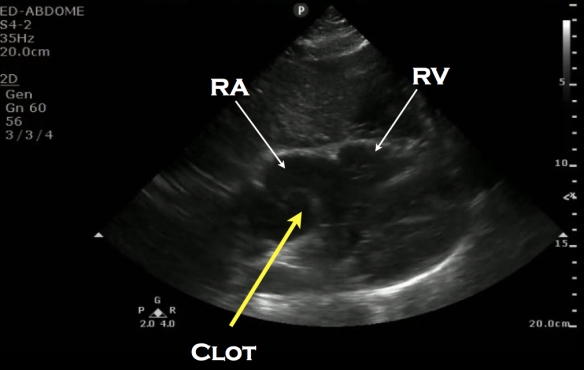
Another, less commonly seen finding would be directly seeing the clot in the right atrium (RA) or in the pulmonary arteries.
Clot in RA:
Clot in pulmonary artery:
Of more practical use are two other sonographic findings: Deep venous thrombosis (DVT) and distal pulmonary infarction.
In a study that included 199 examinations, bedside 2-point compression evaluation of the greater saphenous/femoral vein junction and the popliteal veins of patients with suspected DVT was found to be 100% sensitive and 99% specific for DVT 9. However, it is possible for a patient to present with an acute PE and have a negative DVT, and only about 40-50% of patients with DVT’s will end up having a PE10, 11
DVT on one side diagnosed by noncompressible vein:
More recently, lung ultrasound has been explored for the assessment of a suspected PE. A recent systematic review and meta-analysis by Squizzato et al which included 10 studies and a total of 887 patients found lung ultrasound to have a mean sensitivity of 87% and a mean specificity of 82% for acute PE12. What they looked for in the lung was the presence of triangular, wedge or rounded hypoechoic, pleural based lesions. These lesions are thought to be due to embolic occlusions that resulted in either focal atelectasis with extravasation of blood or focal infarction of the lung parenchyma . However, they state in their publication that “Several methodological drawbacks of the primary studies limit any definite conclusion”.
Lung infarction:
Instead of looking at just one specific sonographic finding for the diagnosis of acute PE, a better method may the use of multi-organ sonography. Recently, Nazerian et al. published a study utilizing multi-organ sonography in the diagnosis of PE. This study used echo, lung and DVT ultrasound to diagnose PE and found that when the three ultrasounds were combined, they yielded a sensitivity of 90%, which was significantly higher than each of the exams by themselves13.
Like any physical exam finding, lab reports or other radiographic assessments, the sonographic analysis of a patient with a suspected pulmonary embolism should be used as part of your diagnostic quiver, and not the silver bullet. Any of the above mentioned ultrasound findings of acute PE can potentially be found in other, non-PE causes of dyspnea. DVT’s can just be DVT’s, RV enlargement can be chronic or from an RV infarction, and subpleural fluid collections can be seen in contusions, pneumonia and cancer. This doesn’t mean not to use it though. Just think about all the other tests we use in the emergency department, such as EKG’s, chest x-rays, troponins, BNP, and the d-dimer. All of these can be abnormal in PE and in non-PE entities.
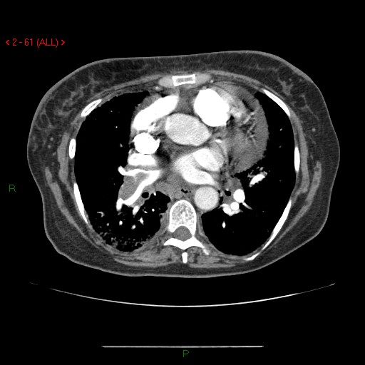
Now back to our patient. She is a 46 year-old female with COPD that had right heart enlargement, which we learned above can be seen in COPD without the presence of a PE. You were unable to get a good subcostal view of the heart to measure the lateral wall, mostly because the patient did not tolerate being laid flat. You move on to the lungs and in the lower right thorax and there you find two hypoechoic, pleural based lesions. Heparin and a CT scan are ordered, and the CT scan shows a large clot located in the right main pulmonary artery.
Here is the CT scan showing the clot:
To see a recent podcast by Ultrasoundpodcast on multi-organ US for PE, go here.
References:
- Otto, Catherine M.. Textbook of clinical echocardiography. 5th ed. Philadelphia, PA: Elsevier/Saunders, 2013. Print. p 247
- Wood KE. Major pulmonary embolism: review of a pathophysiologic approach to the golden hour of hemodynamically significant pulmonary embolism. Chest. 2002;121:877-905
- Jelinek GA, Ingarfield SL, Mountain D, et al. Emergency department diagnosis of pulmonary embolism is associated with significantly reduced mortality: a linked data population study. Emerg Med Australas. 2009;21:269-276
- Goldhaber SZ, Bounamenaux H. Pulmonary embolism and deep vein thrombosis. Lancet 2012:379:1835-46
- Dresden S1, Mitchell P2, Rahimi L2, Leo M2, Rubin-Smith J2, Bibi S2, White L3, Langlois B2, Sullivan A4, Carmody K5 Right ventricular dilatation on bedside echocardiography performed by emergency physicians aids in the diagnosis of pulmonary embolism. Ann Emerg Med. 2014 Jan;63(1):16-24. doi: 10.1016/j.annemergmed.2013.08.016. Epub 2013 Sep 27.
- Rudski LG, Lai WW, Afilalo J, Hua L, Handschumacher MD, Chandrasekaran K et al. Guidelines for the echocardiographic assessment of the right heart in adults: a report from the American Society of Echocardiography endorsed by the European Association of Echocardiography, a registered branch of the European Society of Cardiology, and the Canadian Society of Echocardiography. J Am Soc Echocardiogr. 2010;23:685-713
- McConnell MV, Solomon SD, Rayan ME, Come PC, Goldhaber SZ, Lee RT. Regional right ventricular dysfunction detected by echocardiography in acute pulmonary embolism. Am J Cardiol 1996;78:469e73.
- Casazza F, Bongarzoni A, Capozi A, Agostoni O. Regional right ventricular dysfunction in acute pulmonary embolism and right ventricular infarction. Eur J Echocardiogr. 2005;6:11-4
- Crisp JG, Lovato LM, Jang T. Compression ultrasonography of the lower extremity with portable vascular ultrasonography can accurately detect deep venous thrombosis in the emergency department. Ann Emerg Med. 2010;56:601-610
- Kearon C. Natural history of venous thromboembolism. Circulation. 2003;107:22-30
- Moser KM, Fedullo PF, Littlejohn JK, Crawford R. Frequent asymptomatic pulmonary embolism in patients with deep venous thrombosis. JAMA 1994;271:223-225
- Squizzato A1, Rancan E, Dentali F, Bonzini M, Guasti L, Steidl L, Mathis G, Ageno W. Diagnostic accuracy of lung ultrasound for pulmonary embolism: a systematic review and meta-analysis. J Thromb Haemost. 2013 Jul;11(7):1269-78. doi: 10.1111/jth.12232.
-
Nazerian P, et al. Accuracy of Point-of-Care Multiorgan Ultrasonography for the Diagnosis of Pulmonary Embolism.Chest. 2014 May 1;145(5):950-7. doi: 10.1378/chest.13-1083


Pingback: Subpleural Consolidations in PE – Core Ultrasound
Thanks Jacob for posting such an informative and useful post on echocardiography that too for free. I am a pediatric cardiologist by profession and have been searching all the time on the internet all about echocardiography. I enjoyed a lot reading your post and learnt many new things on this topic. I would like to again thank you for such a nice post. Keep writing for the benefit of readers like us.
Thank you for reading!
Pingback: Subpleural Consolidations in PE | 5 minute sono
Really liked this. Spot on. Nazerian’s LR’s were really useful.
Thanks for the comment! Ill definitely let Jabob know.
Thanks, I did not mean to be so alliterative. I was not sure about his right atrial clot though….might have been my poor technology, but I thought it looked more like one of those congenital incidentals.
from Jacob – Thanks for your comment/feedback. There are definitely congenital incidentals that can cause false positive findings on ultrasound, and being able to identify them in the heat of the moment while managing a crashing patient takes skill and experience. In the case of the RA clot, the most likely etiology would be an actual clot rather than a congenital incidental as this particular patient was crashing and had a positive femoral DVT. The pulmonary artery clot could also be a tricky one, but in this particular patient, the clot in the pulmonary artery was confirmed by CT scan. I’ve labeled the pictures, and hopefully that helps clear things up!
Yes, thanks heaps…I need to learn how to zoom better on my computer.
KB