SonoDoc: A free online POCUS game!

It’s here! SonoDoc: A fun, online game to learn bedside #ultrasound in a case-based way. Learn #POCUS indications, how to’s, integrate into case mgmt, how to use it for next steps/dispo. Play FREE or Stanford CME ($25). HUGE thanks to many folks for authoring cases/beta testing/feedback/support. 5 years in the making, and so happy to share it with you all. Enjoy! http://sonodocgame.com

Screen Shot 2019-02-14 at 10.18.55 PM

SonoApp- Bedside ultrasound for thyroid nodules? – a patient’s question and experience

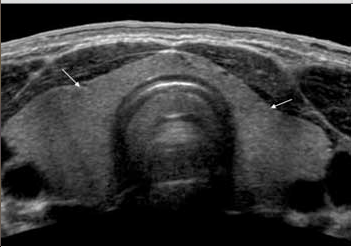
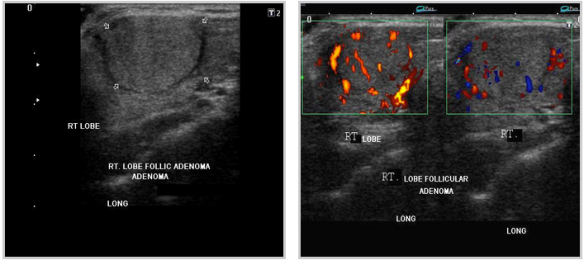
This post involves a personal experience from a free lance writer who asked me whether we (EM or primary care docs) should/can be doing bedside ultrasound for an assessment of the thyroid when we feel something wrong – even if its for the sole purpose of expediting care and biopsy, or alleviating concerns with a negative study.  It was a valid question, and she wrote about her experience below. The thyroid is not a difficult gland to find, nor is it a technically difficult ultrasound to perform – although not a current use for bedside ultrasound, it may start to be given the ease to which nodules can be seen: (for pictures of normal thyroid and abnormal thyroid nodules, see the end of the post  – the pics are not her thyroid)

Point-of-Care Ultrasounds: Can They be Used for Thyroid Nodules?

At a regular physical exam, my doctor discovered a small lump in my neck. She said it was on my thyroid, and referred me to an endocrinologist for further evaluation. At only 19 years old, I was scared and extremely uneducated in regards to thyroid health and diagnoses. My endocrinologist ordered a radiology ultrasound to be done at a later time where I had the technician hovering over me. After the doctor declared I had two nodules, I was scheduled to go back yet again for an ultrasound-guided fine needle aspiration (FNA) to help rule out cancer. This persistent waiting from one appointment to another for small amounts of information were frustrating, and I wondered why my own doctor couldn’t just do the initial ultrasound to visualize the nodules, streamlining the process and expediting my work up.

The day finally arrived for the FNA, and the scene couldn’t be more uncomfortable—and comical at the same time. As the technician tried to gain an image of the right spots with the large wand, the endocrinologist struggled to bypass the size of it in order to gain the samples he needed. On top of that, it was nearly four months from the time of my original physical exam before I found out I had benign nodules. I was told I would need to follow up with a radiology ultrasound every few years.

Point-of-care (POC) ultrasounds, units that are small and possibly hand-held that are increasing in popularity among physicians and emergency doctors, can used by primary care doctors with their assessment. Not only are they portable, but they are easier and more comfortable to use for all parties involved. While not widely publicized or used for thyroid nodules yet, there is potential that POC ultrasounds could aid in diagnosis and follow-up for related patients.

Defining Thyroid Nodules

Thyroid nodules are areas of abnormal cell growths within the thyroid gland. Depending on their size, nodules may form a visible mass in the neck. Some can also cause a goiter, or enlarged thyroid gland. The majority of thyroid nodules are not cancerous. However, an ultrasound and FNA can rule this out. These nodules may go undetected for years. Others can coexist with other related issues, such as hypothyroidism and hyperthyroidism. Thyroid nodules can also become painful to the touch and when you swallow.

Unless you can feel a lump in your thyroid gland, these types of nodules are first detected through a physical exam of the neck. Conventional radiology ultrasounds are currently used as a follow-up to locate the nodules and rule out any potential problems.

Role of POC Ultrasounds

On a larger scale, the increased use of POC ultrasounds can help the health of patients. It can even help save lives: although rare, thyroid cancer can be deadly if not caught early. Most cases of thyroid nodules and masses occur in older patients, some of whom may not be able to make it to radiology offices for a traditional ultrasound. By employing the use of a POC ultrasound, a physician can quicken the diagnosis and make the process more comfortable for patients. The fact is that once patients leave their primary care physicians’ offices, it is up to them to follow up on referrals to ultrasound appointments. Having a POC device handy ensures that a patient won’t miss these crucial diagnoses.

Having an ultrasound for thyroid nodules is easier with POC ultrasounds. Since the area of investigation is small, this approach makes the most sense when considering comfort for both doctor and patient.

Another benefit is the cost. POC ultrasounds cost providers less money, and it may be more affordable to patients. With the rising costs of healthcare, more and more patients are missing out on critical exams. By making thyroid scans more affordable, patients may be less likely to skip out on their appointments. The downside is that POC ultrasounds are not used for thyroid nodules yet. By pushing for their use in all areas of healthcare, POC may ease the ultrasound experience and even save lives.

Resources

Kristeen Cherney

Author Bio: Kristeen Cherney is a freelance health and lifestyle writer who also has a certificate in nutrition. Her work has been published on numerous health-related websites.

Pics from aium.org – NORMAL ADULT THYROID

normal thyroid

Pics form ultrasound-images.com :

ADENOMAS

thyroid_adenomatous nodules thyroid_follicular adenoma

FOLLICULAR ADENOMAthyroid_follicular adenomas

PAPILLARY CARCINOMAthyroid_papillary carcinoma


SonoTutorial: Ultrasound guided Suprapubic bladder aspiration – in NEJM #FOAMus

Ok, this is awesome. I know you are likely thinking that i am talking about ultrasound and how it helps with bladder aspiration, and although i do think that is awesome, it’s not that which makes me smile right now. It is that it is highlighted in the New England Journal of Medicine – oh yes, that’s right. You’ll need to be a subscriber to see the video, but you can see the front page of the article here. Hooray for the NEJM on highlighting ultrasound in this important application for our patients!


SonoStudy: Trends in Radiology orders over last decade – effect of POCUS? #FOAMus

I am going to say a statement that is going to be shocking to some of you: There has been a decline in CT use by the emergency department over the last 2 decades, according to Raja et al. Can you believe it? I couldnt either. I previously thought, now that CT is so easy to get, of course everyone is ordering them more. There have been several studies showing an increase in ionizing radiation exposure over the last decade, so the results are a bit confusing. Im sure when compared to 20+ years ago, we are ordering more in total, but the trend may be that we are declining in ordering. Well, not only has Raja came to this conclusion, but when seeing how the FAST scan has affected abdominal CT scan orders, then it makes me wonder if point of care ultrasound (POCUS) is one of the main reasons for this trend. Yeah, I know, it’s a stretch, but I cannot imagine it isnt a factor, along with ALARA, and other discussions on radiation exposure.

In a study by Sheng et al – which includes some of my heroes, Drs. Vickie Noble and Andrew Liteplo – they looked at the trend of abdominal CT orders in adult trauma patients at their institution. Could it be that bedside ultrasound has effected CT orders everywhere? Their abstract is below:

Objective. We sought to describe the trend in abdominal CT use in adult trauma patients after a point-of-care emergency ultrasound program was introduced. We hypothesized that abdominal CT use would decrease as FAST use increased. Methods. We performed a retrospective study of 19940 consecutive trauma patients over the age of 18 admitted to our level one trauma center from 2002 through 2011. Data was collected retrospectively and recorded in a trauma registry. We plotted the rate of FAST and abdominal CT utilization over time. Head CT was used as a surrogate for overall CT utilization rates during the study period. Results. Use of FAST increased by an average of 2.3% (95% CI 2.1 to 2.5, P < 0.01) while abdominal CT use decreased by the same rate annually. The percentage of patients who received FAST as the sole imaging modality for the abdomen rose from 2.0% to 21.9% while those who only received an abdominal CT dropped from 21.7% to 2.3%. Conclusions. Abdominal CT use in our cohort declined while FAST utilization grew in the last decade. The rising use of FAST may have played a role in the reduction of abdominal CT performed as decline in CT utilization appears contrary to overall trends.

SonoCredentialing: ACGME ultrasound milestones summarized by @Takeokun

@Takeokun is not only one of the authors for the CORD article on this topic, but also has summarized the ACGME milestones’ ultrasound portion quite nicely on his site. It is well deserving of mention –

Go here for the full post.

Some Downloadables from his post:

  1. Core vs Advanced Emergency Ultrasound for Residents
  2. Assessment methods with skills tested, limitations, and associated milestones
  3. CORD US-SDOT Forms
  4. New Innovations version of the JMTF US milestone evaluation form

SonoGlobalHealth – Ultrasound in Global Health resources available @AIUM_Ultrasound

Screen Shot 2014-07-23 at 11.14.23 AM

For those of us who travel, teach, and spread the gospel of ‘sound – we look for resources that may help in knowing the conversation for this topic. AIUM has a page that could help in your needs, as well as see photos of trips that their physicians have undertaken for ultrasound teaching and advancement.

Ive posted on other groups that have gone all over the world, in addition to my own experiences – including TO-theWorld who enhanced maternal education and minimizing MTFT of HIV.

For other resources, that are free for ultrasound in global health:

Partners in Health

World Health organization

Here’e a video that discusses ultrasound in global health, with a cameo photo of our own Dr. Jessica Ngo using it during Stanford’s emergency response to Haiti after the disaster with our SEMPER team.

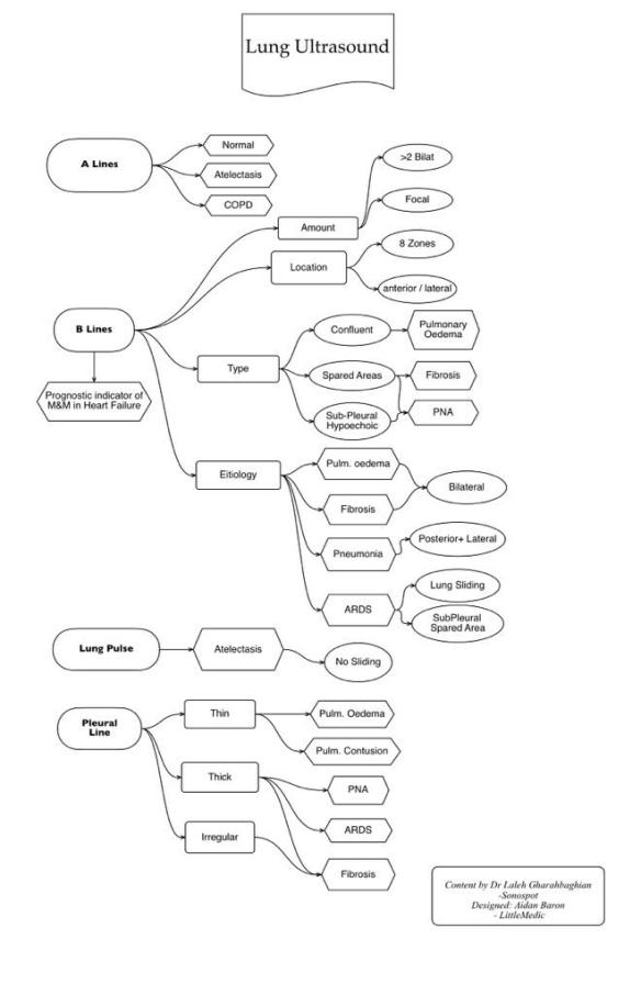
SonoFlowCharts – The Lung Ultrasound Flowchart – A lines & B lines #FOAMed #FOAMus

When I teach my fellows/residents/students in the Ultrasound office, there is a white board behind me, which I’ll use to draw, write, and place arrows linking one thing to another – it doesnt ever look that great in its final form. Then @LittleMedic – an awesome blogger/student in Sydney Australia –  took the pic I posted of my white board scratches and made it read-able! Oh, if you only knew what the board looked like, you would thank him with all your heart, as I do now. Lung US flow chart

SonoGuidelines : AIUM guidelines for cleaning probes (external and internal) #FOAMus

Update: ACEP has written guidelines as of June 2018 – find it here

In an official statement by the American Institute of Ultrasound in Medicine (AIUM), they updated their guidelines on cleaning probes: Read below:

“The purpose of this document is to provide guidance regarding the cleaning and preparation of external and internal ultrasound probes. Some manufacturers use the term “transducers” or “imaging arrays.”

Medical instruments fall into different categories with respect to their potential for pathogen transmission. The most critical instruments are those that are intended to penetrate skin or mucous membranes. These require sterilization. Less critical instruments (often called “semicritical” instruments) that simply come into contact with mucous membranes, such as fiber-optic endoscopes, require high-level disinfection rather than sterilization. “Noncritical” devices come into contact with intact skin but not mucous membranes.

External probes that only come into contact with clean, intact skin are considered noncritical devices and require cleaning after every use as described below.

All internal probes should be covered with a single-use barrier. If condoms are used as barriers, they should be nonlubricated and nonmedicated. Although internal ultrasound probes are routinely protected by single-use disposable probe covers, leakage rates of 0.9% to 2% for condoms and 8% to 81% for commercial probe covers have been observed in recent studies (Rutala and Weber, 2011). These probes are therefore classified as semicritical devices.

Note: Practitioners should be aware that condoms have been shown to be less prone to leakage than commercial probe covers and have a 6-fold enhanced acceptable quality level (AQL) when compared to standard examination gloves. They have an AQL equal to that of surgical gloves. Users should be aware of latex sensitivity issues and have non-latex-containing barriers available.

For maximum safety, one should therefore perform high-level disinfection of the probe between each use and use a probe cover or condom as an aid to keep the probe clean. For the purpose of this document, “internal probes” refer to all vaginal, rectal, and transesophageal probes, as well as intraoperative probes and all probes that are in contact with bodily fluids/blood or have a remote chance to be in contact with dry/cracked skin and body fluids, including blood.

Definitions

All cleaning, disinfection, and sterilization represent a statistical reduction in the number of microbes present on a surface rather than their complete elimination. Meticulous cleaning of the instrument is the key to an initial reduction of the microbial/organic load by at least 99%. This cleaning is followed by a disinfecting procedure to ensure a high degree of protection from infectious disease transmission, even if a disposable barrier covers the instrument during use.

According to the Centers for Disease Control and Prevention (CDC) “Guideline for Disinfection and Sterilization in Healthcare Facilities” (2008):

Cleaning is the removal of visible soil (eg, organic and inorganic material) from objects and surfaces and normally is accomplished manually or mechanically using water with detergents or enzymatic products. Thorough cleaning is essential before high-level disinfection and sterilization because inorganic and organic material that remains on the surfaces of instruments interfere with the effectiveness of these processes.”

Disinfection describes a process that eliminates many or all pathogenic microorganisms, except bacterial spores.”

Low-Level Disinfection—Destruction of most bacteria, some viruses, and some fungi. Low-level disinfection will not necessarily inactivate Mycobacterium tuberculosis or bacterial spores.

Mid-Level Disinfection—Inactivation of M Tuberculosis, bacteria, most viruses, most fungi, and some bacterial spores.

High-Level Disinfection—Destruction/removal of all microorganisms except bacterial spores.

Sterilization describes a process that destroys or eliminates all forms of microbial life and is carried out in healthcare facilities by physical or chemical methods. Steam under pressure, dry heat, ethylene oxide (EtO) gas, hydrogen peroxide gas plasma, and liquid chemicals are the principal sterilizing agents used in health-care facilities. . . . When chemicals are used to destroy all forms of microbiologic life, they can be called chemical sterilants. These same germicides used for shorter exposure periods also can be part of the disinfection process (ie, high-level disinfection).”

The following specific recommendations are made for the cleaning and preparation of all ultrasound probes. Users should also review the CDC document on sterilization and disinfection of medical devices to be certain that their procedures conform to the CDC principles for disinfection of patient care equipment.

1. Cleaning—Transducers should be cleaned after each examination with soap and water or quaternary ammonium (a low-level disinfectant) sprays or wipes. The probes must be disconnected from the ultrasound scanner for anything more than wiping or spray cleaning. After removal of the probe cover (when applicable), use running water to remove any residual gel or debris from the probe. Use a damp gauze pad or other soft cloth and a small amount of mild nonabrasive liquid soap (household dish-washing liquid is ideal) to thoroughly cleanse the probe. Consider the use of a small brush, especially for crevices and areas of angulation, depending on the design of the particular probe. Rinse the probe thoroughly with running water, and then dry the probe with a soft cloth or paper towel.

2. Disinfection—As noted above, all internal probes (eg, vaginal, rectal, and transesophageal probes) as well as intraoperative probes require high-level disinfection before they can be used on another patient.

For the protection of the patient and the health care worker, all internal examinations should be performed with the operator properly gloved throughout the procedure. As the probe cover is removed, care should be taken not to contaminate the probe with secretions from the patient. At the completion of the procedure, hands should be thoroughly washed with soap and water. Gloves should be used to remove the probe cover and to clean the probe as described above.

Note: An obvious disruption in condom integrity does not require modification of this protocol. Because of the potential disruption of the barrier sheath, high-level disinfection with chemical agents is necessary. The following guidelines take into account possible probe contamination due to a disruption in the barrier sheath.

After removal of the probe cover, clean the transducer as described above. Cleaning with a detergent/water solution as described above is important as the first step in proper disinfection, since chemical disinfectants act more rapidly on clean and dry surfaces. Wet surfaces dilute the disinfectant.

High-level liquid disinfection is required to ensure further statistical reduction in the microbial load. Examples of such high-level disinfectants are listed in Table 1. A complete list of US Food and Drug Administration (FDA)-cleared liquid sterilants and high-level disinfectants is available at http://www.fda.gov/MedicalDevices/Safety/AlertsandNotices/ucm194429.htm, and other agents are under investigation.

To achieve high-level disinfection, the practice must meet or exceed the listed “High-Level Disinfectant Contact Conditions” specified for each product. Users should be aware that not all approved disinfectants on this list are safe for all ultrasound probes.

The CDC recommends environmental infection control in the case of Clostridium difficile, consisting of “meticulous cleaning followed by disinfection using hypochlorite-based germicides as appropriate” (CDC, 2008). The current introduction and initial marketing of a hydrogen peroxide nanodroplet emulsion might provide an effective high-level disinfectant without toxicity.

Table 1. Sterilants and High-Level Disinfectants Listed by the FDA

Name Composition/Action
Glutaraldehyde Organic compound (CH2(CH2CHO)2)
Induces cell death by cross-linking cellular proteins; usually used alone or mixed with formaldehyde
Hydrogen peroxide Inorganic compound (H2O2)
Antiseptic and antibacterial; a very strong oxidizer with oxidation potential of 1.8 V
Peracetic acid Organic compound (CH3CO3H)
Antimicrobial agent (high oxidization potential)
Ortho-Phthalaldehyde Organic compound (C6H4(CHO)2)
strong binding to outer cell wall of contaminant organisms
Hypochlorite/hypochlorous acid inorganic compound (HClO)
Myeloperoxidase-mediated peroxidation of chloride ions
Phenol/phenolate Organic compound (C6H5OH)
Antiseptic
Hibidil Chlorhexidine gluconate (C22H30Cl2N10)
Chemical antiseptic

The Occupational Safety and Health Administration as well as the Joint Commission (Environment of Care Standard IC 02.02.01 EP 9) have issued guidelines for exposure to chemical agents, which might be used for ultrasound probe cleaning. Before selecting a high-level disinfectant, users should request the Material Safety Data Sheet for the product and make sure that their facility is able to meet the necessary conditions to minimize exposure (via inhalation, ingestion, or contact through skin/eyes) to potentially dangerous substances. Proper ventilation, a positive-pressure local environment, and the use of personal protective devices (eg, gloves and face/eye protection) may be required.

Immersion of probes in fluids requires attention to the individual device’s ability to be submerged. Although some scan heads as well as large portions of the cable may safely be immersed up to the connector to the ultrasound scanner, only the scan heads of others may be submerged. Some manufacturers also note that the crystals of the array may be damaged if, instead of suspending the probe in the disinfectant, it rests on the bottom of the container. Before selecting a method of disinfection, consult the instrument manufacturer regarding the compatibility of the to-be-used agent with the probes. Relevant information is available online and in device manuals. Additionally, not all probes can be cleaned with the same cleaning agents. Although some agents are compatible with all probes of a given manufacturer, others must be limited to a subset of probes.

After soaking the probe in an approved disinfectant for the specified time, the probe should be thoroughly rinsed (especially to remove traces of toxic disinfectants in the case of ortho-phthalaldehyde) and dried.

Summary

Adequate probe preparation is mandatory. The level of preparation depends on the type of examination performed. Routine high-level disinfection of internal probes between patients is mandatory, plus the use of a high-quality single-use probe cover during each examination is required to properly protect patients from infection. It would be reassuring for the user to be able to consult manufacturer’s instructions, particularly those that have been validated by the manufacturer for sterilizing devices. Preparation of external probes between patients is less critical and reduced to a low-level disinfection process. For all chemical disinfectants, precautions must be taken to protect workers and patients from the toxicity of the disinfectant.

The AIUM does not endorse or promote any specific commercial products. It is the responsibility of each entity to follow the manufacturer’s guidelines, law, and regulations.

Suggested Reading

  1. Amis S, Ruddy M, Kibbler CC, Economides DL, MacLean AB. Assessment of condoms as probe covers for transvaginal sonography. J Clin Ultrasound 2000; 28:295-298.
  2. Bloc S, Garnier T, Bounhiol C, et al. Ultrasound-Guided Regional Anaesthesia: An Effective Method for Cleaning the Probes. Quincy-Sous-Sénart, France: Service d’Anesthésie, Hôpital Privé Claude-Galien; 2010.
  3. Centers for Disease Control and Prevention, Hospital Infections Program. National Nosocomial Infections Surveillance (NNIS) report, data summary from October 1986-April 1996: a report from the NNIS system. Am J Infect Control 1996; 24:380-388.
  4. Frazee BW, Fahimi J, Lambert L, Nagdev A. Emergency department ultrasonographic probe contamination and experimental model of probe disinfection. Ann Emerg Med 2011; 58:56-63.
  5. Hignett M, Claman P. High rates of perforation are found in endovaginal ultrasound probe covers before and after oocyte retrieval for in vitro fertilization-embryo transfer. J Assist Reprod Genet 1995; 12:606-609.
  6. Kac G, Podglajen I, Si-Mohamed A, Rodi A, Grataloup C, Meyer G. Evaluation of Ultraviolet C for Disinfection of Endocavitary Ultrasound Transducers Persistently Contaminated Despite Probe Covers. Paris, France: Hygiène Hospitalière; 2010
  7. Koibuchi H, Fujii Y, Kotani K, et al. Degradation of ultrasound probes caused by disinfection with alcohol. J Med Ultrason 2011; 38:97-100.
  8. Milki AA, Fisch JD. Vaginal ultrasound probe cover leakage: implications for patient care. Fertil Steril 1998; 69:409-411.
  9. Mirza WA, Imam SH, Kharal MS, et al. Cleaning methods for ultrasound probes. J Coll Physicians Surg Pak 2008; 18:286-289.
  10. Muradali D, Gold WL, Phillips A, Wilson S. Can ultrasound probes and coupling gel be a source of nosocomial infection in patients undergoing sonography? An in vivo and in vitro study. AJR Am J Roentgenol 1995; 164:1521-1524.
  11. Rooks VJ, Yancey MK, Elg SA, Brueske L. Comparison of probe sheaths for endovaginal sonography. Obstet Gynecol 1996; 87:27-29.
  12. Rutala WA, Weber DJ. Sterilization, high-level disinfection, and environmental cleaning. Infect Dis Clin North Am 2011; 25:45-76.
  13. Whitehead EJ, Thompson JF, Lewis DR. Contamination and decontamination of Doppler probes. Ann R Coll Surg Engl 2006; 88:479-481.

Related Websites

  1. US Food and Drug Administration. Cleared liquid chemical sterilants/high-level disinfectants US FDA website; March 03, 2010. http://www.fda.gov/MedicalDevices/Safety/AlertsandNotices/ucm194429.htm.
  2. Centers for Disease Control and Prevention. Guideline for disinfection and sterilization in healthcare facilities, 2008. Centers for Disease Control and Prevention website; 2008. http://www.cdc.gov/hicpac/pdf/guidelines/Disinfection_Nov_2008.pdf.
  3. Occupational Safety and Health Administration. Hospital eTool: clinical services. Occupational Safety and Health Administration website. http://www.osha.gov/SLTC/etools/hospital/clinical/pt/pt.html.

SonoCase: 46yo shortness of breath – guest post by Jacob Avila @UltrasoundMD #FOAMed

Our newest guest post is by one of the best emergency medicine resident educators I know – of course, you dont want to miss his educational pearls on twitter too – Dr. Jacob Avila. He discusses a case that illustrates how bedside ultrasound can help in your unexplained short of breath patient, and even cancel that triage bias that your attending can do to sway you away from the truth. Let’s give it to Dr. Avila for highlighting (with a great literature review) how ultrasound can help you too. Here it is – enjoy! (note: not all images were of this patient, but were taken from other resources)

“You arrive to the emergency department for your first night shift of the month, and as you place your bag on the desk, the attending walks towards you with a chart in his hand. “Do you mind seeing this patient? It’s a COPD’er with dyspnea.  It’s probably just a COPD exacerbation.”  You look at the chart and see that it’s a 46 year old female with shortness of breath.  As you walk into the room, you notice the patient appears slightly pale, is afebrile, has an O2 saturation of 91% and is tachycardic in the 110’s with a blood pressure of 105/76, temperature of 98.5° and respiratory rate of 26.  While taking the history, you note that the patient is a smoker and recently returned from a 12 hour car ride to see relatives.  Suspecting that this may be something other than simple COPD exacerbation, you grab your ultrasound machine and start with the cardiac echo (as described in the RADiUS protocol) and are able to get the following image:

This apical 4-chamber view shows severe right heart dilation, defined as a RV:LV ratio >1.  However, you remember that the patient has a history of COPD, and chronic pulmonary hypertension can cause chronic right ventricular dilation1.  At that moment, the patient becomes hypotensive with a systolic blood pressure in the 70’s and develops severe respiratory distress. What should we do?

Early diagnosis of a pulmonary embolism (PE) is exceedingly important, as two thirds of patients with mortality associated with a PE die within the first hour of their presentation2, and intuitively, those who are treated earlier generally have a better prognosis 3.  The definitive diagnosis of a PE requires the use of a CT scanner 4, but in a patient who is unstable, like this one, that isn’t an option. Looking at the right ventricular to left ventricular ratio is a maneuver that can rapidly change your differential diagnosis or confirm what you previously suspected. A recent study by Dresden et al found right ventricular dilatation identified by emergency physicians had a specificity of 98% for a PE.  That number is impressive, but when you look at the methodology section of the publication, only 10% of the patients they included had coexisting COPD, and all of the false positives in the study were in patients with COPD5.   One  technique that may help differentiate between chronic and acute dilation is looking at the RV free wall in the subxiphoid view while in end diastole. A free wall size >0.5 cm is more likely to be chronic RV dilation6i. However, this view is not always possible in all patients.  Another echo sign you could look for is the McConnell sign (apical winking of the right ventricle during systole), which previously was reported to have an impressive 94% specificity and 77% sensitivity for an acute PE7l, but a subsequent and larger study found the McConnell sign to be only 70% sensitive and 33% specific for a PE8. Take a look at what the McConnell sign looks like”


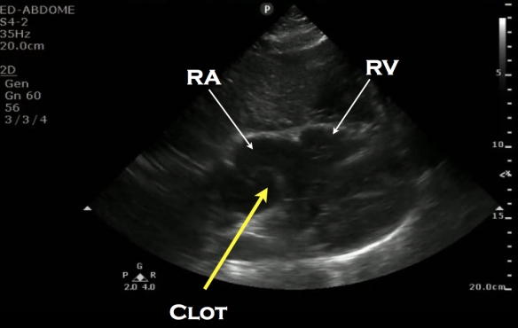
Another, less commonly seen finding would be directly seeing the clot in the right atrium (RA) or in the pulmonary arteries.

Clot in RA:

RA clot labeled

Clot in pulmonary artery:

Pulm clot labeled

Of more practical use are two other sonographic findings: Deep venous thrombosis (DVT) and distal pulmonary infarction.

In a study that included 199 examinations, bedside 2-point compression evaluation of the greater saphenous/femoral vein junction and the popliteal veins of patients with suspected DVT was found to be 100% sensitive and 99% specific for DVT 9.   However, it is possible for a patient to present with an acute PE and have a negative DVT, and only about 40-50% of patients with DVT’s will end up having a PE10, 11

DVT on one side diagnosed by noncompressible vein:

More recently, lung ultrasound has been explored for the assessment of a suspected PE. A recent systematic review and meta-analysis by Squizzato et al which included 10 studies and a total of 887 patients found lung ultrasound to have a mean sensitivity of 87% and a mean specificity of 82% for acute PE12.  What they looked for in the lung was the presence of triangular, wedge or rounded hypoechoic, pleural based lesions.  These lesions are thought to be due to embolic occlusions that resulted in either focal atelectasis with extravasation of blood or focal infarction of the lung parenchyma . However, they state in their publication that “Several methodological drawbacks of the primary studies limit any definite conclusion”.

Lung infarction:


Instead of looking at just one specific sonographic finding for the diagnosis of acute PE, a better method may the use of multi-organ sonography.  Recently, Nazerian et al. published a study utilizing multi-organ sonography in the diagnosis of PE.  This study used echo, lung and DVT ultrasound to diagnose PE and found that when the three ultrasounds were combined, they yielded a sensitivity of 90%, which was significantly higher than each of the exams by themselves13.

Like any physical exam finding, lab reports or other radiographic assessments, the sonographic analysis of a patient with a suspected pulmonary embolism should be used as part of your diagnostic quiver, and not the silver bullet.  Any of the above mentioned ultrasound findings of acute PE can potentially be found in other,  non-PE causes of dyspnea.  DVT’s can just be DVT’s, RV enlargement can be chronic or from an RV infarction, and subpleural fluid collections can be seen in contusions, pneumonia  and cancer.  This doesn’t mean not to use it though.  Just think about all the other tests we use in the emergency department, such as EKG’s, chest x-rays, troponins, BNP, and the d-dimer.  All of these can be abnormal in PE and in non-PE entities.

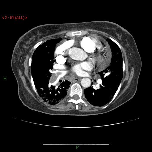
Now back to our patient.  She is a 46 year-old female with COPD that had right heart enlargement, which we learned above can be  seen in COPD without the presence of a PE.  You were unable to get a good subcostal view of the heart to measure the lateral wall, mostly because the  patient did not tolerate being laid flat.  You move on to the lungs and in the lower right thorax and there you find two hypoechoic, pleural based lesions.  Heparin and a CT scan are ordered, and the CT scan shows a large clot located in the right main pulmonary artery.

Here is the CT scan showing the clot:

Avila_Clot in pulmonary artery CT

To see a recent podcast by Ultrasoundpodcast on multi-organ US for PE, go here.

References:

  1. Otto, Catherine M.. Textbook of clinical echocardiography. 5th ed. Philadelphia, PA: Elsevier/Saunders, 2013. Print. p 247
  2. Wood KE. Major pulmonary embolism: review of a pathophysiologic approach to the golden hour of hemodynamically significant pulmonary embolism. Chest. 2002;121:877-905
  3. Jelinek GA, Ingarfield SL, Mountain D, et al. Emergency department diagnosis of pulmonary embolism is associated with significantly reduced mortality: a linked data population study. Emerg Med Australas. 2009;21:269-276
  4. Goldhaber SZ, Bounamenaux H. Pulmonary embolism and deep vein thrombosis. Lancet 2012:379:1835-46
  5. Dresden S1, Mitchell P2, Rahimi L2, Leo M2, Rubin-Smith J2, Bibi S2, White L3, Langlois B2, Sullivan A4, Carmody K5 Right ventricular dilatation on bedside echocardiography performed by emergency physicians aids in the diagnosis of pulmonary embolism. Ann Emerg Med. 2014 Jan;63(1):16-24. doi: 10.1016/j.annemergmed.2013.08.016. Epub 2013 Sep 27.
  6. Rudski LG, Lai WW, Afilalo J, Hua L, Handschumacher MD, Chandrasekaran K et al. Guidelines for the echocardiographic assessment of the right heart in adults: a report from the American Society of Echocardiography endorsed by the European Association of Echocardiography, a registered branch of the European Society of Cardiology, and the Canadian Society of Echocardiography. J Am Soc Echocardiogr. 2010;23:685-713
  7. McConnell MV, Solomon SD, Rayan ME, Come PC, Goldhaber SZ, Lee RT. Regional right ventricular dysfunction detected by echocardiography in acute pulmonary embolism. Am J Cardiol 1996;78:469e73.
  8. Casazza F, Bongarzoni A, Capozi A, Agostoni O. Regional right ventricular dysfunction in acute pulmonary embolism and right ventricular infarction. Eur J Echocardiogr. 2005;6:11-4
  9. Crisp JG, Lovato LM, Jang T. Compression ultrasonography of the lower extremity with portable vascular ultrasonography can accurately detect deep venous thrombosis in the emergency department. Ann Emerg Med. 2010;56:601-610
  10. Kearon C. Natural history of venous thromboembolism. Circulation. 2003;107:22-30
  11. Moser KM, Fedullo PF, Littlejohn JK, Crawford R. Frequent asymptomatic pulmonary embolism in patients with deep venous thrombosis. JAMA 1994;271:223-225
  12. Squizzato A1, Rancan E, Dentali F, Bonzini M, Guasti L, Steidl L, Mathis G, Ageno W. Diagnostic accuracy of lung ultrasound for pulmonary embolism: a systematic review and meta-analysis. J Thromb Haemost. 2013 Jul;11(7):1269-78. doi: 10.1111/jth.12232.
  13. Nazerian P, et al. Accuracy of Point-of-Care Multiorgan Ultrasonography for the Diagnosis of Pulmonary Embolism.Chest. 2014 May 1;145(5):950-7. doi: 10.1378/chest.13-1083

SonoStudy: Appendicitis diagnosed by TransVaginal Ultrasound #FOAMed #FOAMus

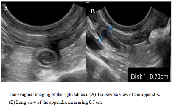
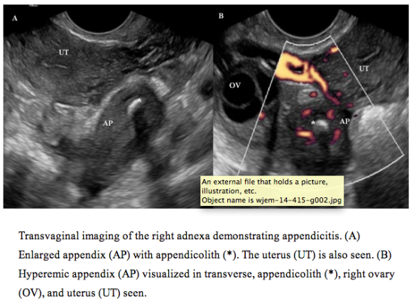
In a publication in WestJEM by Bramante, Raio and team, they discuss two cases of appendicitis found on trans-vaginal ultrasound. Now, this is something that I have been told can happen, but there are few studies on it. It makes sense. First off, the trans-vaginal probe is high frequency, there is an empty bladder and there is a regional evaluation of the pelvis. The appendix can lie low and be visualized, and diagnosed with acute appendicitis. Raio also published a study on this in Emergency Medicine International. They studied 224 females with right lower quadrant pain suspicious for appendicitis excluding those pregnant or under 16yrs old. They had 27 with a positive ultrasound for gyn pathology and 55 had appendicitis per OR report. Of course they wondered if they should have looked for the appendix too! Other studies have shown that with both a transabdominal and a transvaginal ultrasound, you can improve accuracy in appendicitis diagnoses, but that wasnt necessarily to look for the appendix. The Journal of Ultrasound in Medicine published in 2006 about a case of transvaginal US and appendicitis very nicely too. The OB literature also stated how some of their cases of pelvic pain had appendicitis seen on both transabdominal and transvaginal ultrasound.

Ok, back to the case reports in WestJEM by Bramante et al. The 2 cases showed the evaluation and pretty obvious sonographic images seen with acute appendicitis with transvaginal ultrasound after an equivocal transabdominal ultrasound:

Screen Shot 2014-05-23 at 1.38.05 PM Screen Shot 2014-05-23 at 1.38.12 PM