SonoNews: Robotic arm allows remote real-time ultrasound scans -made by French engineers

Now, this is pretty cool. Imagine doing an ultrasound on someone who is not right where you are, but could be in another hospital, or maybe even in another country? These french engineers has made something that is almost to that level. Read below for the EDMT medical imaging article:

Screen Shot 2014-05-15 at 9.19.54 AM

“In a breakthrough that might expand the frontiers of ultrasound examinations, a French company has developed the world’s first industrial-scale remote ultrasound system with a robot arm.

Introduced for radiography, remote imaging is now also used for ultrasound, which accounts for one third of medical imaging procedures, making it the most common such procedure in medicine. Today, ultrasound is the most efficacious of all diagnostic procedures, and on average costs half as much as CT scanning. However, medical exams during pregnancy and detailed investigations of organs, blood vessels, ligaments and the heart require the presence of a highly trained physician at the patient’s bedside as well as hands-on operation of the ultrasound device and interpretation of the resulting images.

Remote medical image scanning using a robot arm that functions as a genuine extension of the physician’s hand was invented by the head of the Department of Nuclear Medicine and Ultrasound at Tours University, Philippe Arbeille, as an outgrowth of manned space flight research that is supported by France’s CNES and by the ESA.

“The main challenge is to resolve the problem of synchronous video transmission. Also, the operator needs to be able to place his hand on the patient virtually,” notes AdEchoTech CEO Éric Lefebvre. The French company recently invested four years and one million Euros in the development of the world’s first industrial-scale remote ultrasound system. AdEchoTech has four patents pending for its Melody robot, two of them with Orleans University’s PRISME mechanical engineering research center.

AdEchoTech’s Melody workstation consists of two elements. For the medical practitioner, the workstation comprises a control box, a mini-console and a virtual probe that remotely controls the ultrasound probe arm. And on the patient side, the operator controls the probe arm remotely. The ultrasound probe, connected to the ultrasound device, is attached to the extremity of the robot arm, whose load is lightened by the arm’s carrier base while at the same time making the arm very easy to manipulate. Thanks to its modularity, the solution is readily adaptable to any given technical environment. Particularly, since operator and patient stations can be operated via satellite, as well as fiber optic 3G+ and 4G hookups. In this regard, the system is compatible with most commercially available ultrasound and video conferencing systems.

“Our R&D also focused on compression algorithms, so as to allow for the communication of information concerning the transmission of ultrasound images, as well as for control of the robot arm and for the realization of video conferences in real time – and all of this in a merely 2 Mbit/s symmetrical bandwidth,” says an AdEchoTech engineer.

The Melody robot, which obtained the CE mark in 2012, allows for 90% concordance with conventional ultrasound, and is useful for solving the growing problems entailed by the performance of ultrasound examinations on islands, in mountainous regions, and on ships or oil drilling platforms, in military zones, for repatriation medicine, and in prisons. Ten Melody stations are already in operation in French hospitals, where they are used for the following procedures: pelvic and abdominal emergencies (investigations of the hepatobiliary system, the urinary system, the abdominal aorta and the pancreas) and examinations during pregnancy. In such settings, the system helps to avoid lengthy trips for pregnant women to major hospitals.

“A second generation of patented Melody robots will benefit from two additional degrees of movement freedom, which will allow for the performance of remote cardiac ultrasound examinations,” says Lefebvre. A third generation of Melody robots, which is expected to be available in 2017, will carry out remote robot-assisted ultrasound for punctures and certain surgical procedures.

By: Bernard Banga, MD Report, France

SonoNews: New Guidelines- reduce risk of premature diagnosis of non-viable pregnancy #FOAMed

In a meeting of 15 members of the radiology, Ob/Gyn, and emergency medicine communities, new criteria were set that was published in NEJM Oct 2013 so that we dont prematurely state that a pregnancy is non-viable. This is pretty important, and a subject that I posted about earlier as well when discussing the usefulness (…or useless ness) of the beta hCG. Can you imagine what was done, and I remember this algorithm – you have a patient with 1st trimester pain or vaginal bleeding, no IUP seen on US, low beta Hcg, and OB was called and the patient was given methotrexate??? Well, there have been cases where those patients actually had a viable IUP that showed up a week later… and then the lawsuit happens….scary stuff. It’s different now where we dont care too much about the beta hCG, or whether there is not an IUP, but whether we see anything around the ovary….and even then, very close follow up and rechecks may be considered. Below is the Eurekalert and the AuntMinnie articles on it too:

Medical experts recommend steps to reduce risk of inadvertent harm to potentially normal pregnancies

New criteria aim to prevent misdiagnoses of nonviable pregnancies

A panel of 15 medical experts from the fields of radiology, obstetrics-gynecology and emergency medicine, convened by the Society of Radiologists in Ultrasound (SRU), has recommended new criteria for use of ultrasonography in determining when a first trimester pregnancy is nonviable (has no chance of progressing and resulting in a live-born baby). These new diagnostic thresholds, published Oct. 10 in the New England Journal of Medicine, would help to avoid the possibility of physicians causing inadvertent harm to a potentially normal pregnancy.

“When a doctor tells a woman that her pregnancy has no chance of proceeding, he or she should be absolutely certain of being correct. Our recommendations are based on the latest medical knowledge with input from a variety of medical specialties. We urge providers to familiarize themselves with these recommendations and factor them into their clinical decision-making,” said Peter M. Doubilet, MD, PhD, of Brigham and Women’s Hospital and Harvard Medical School in Boston, the report’s lead author.

Among the key points made by the expert panel:

  • Until recently, a pregnancy was diagnosed as nonviable if ultrasound showed an embryo measuring at least five millimeters without a heartbeat. The new standards raise that size to seven millimeters
  • The standard for nonviability based on the size of a gestational sac without an embryo should be raised from 16 to 25 millimeters
  • The commonly used “discriminatory level” of the pregnancy blood test is not reliable for excluding a viable pregnancy

The panel also cautioned physicians against taking any action that could damage an intrauterine pregnancy based on a single blood test, if the ultrasound findings are inconclusive and the woman is in stable condition.

Kurt T. Barnhart, MD, MSCE, an obstetrician-gynecologist at the Perelman School of Medicine at the University of Pennsylvania and a member of the SRU Multispecialty Panel, added, “With improvement in ultrasound technology, we are able to detect and visualize pregnancies at a very early age. These guidelines represent a consensus that will balance the use of ultrasound and the time needed to ensure that an early pregnancy is not falsely diagnosed as nonviable. There should be no rush to diagnose a miscarriage; more time and more information will improve accuracy and hopefully eliminate misdiagnosis.”

Michael Blaivas, MD, an emergency medicine physician affiliated with the University of South Carolina and one of the panelists, emphasized that “These are critical guidelines and will help all physicians involved in the care of the emergency patient. They represent an up-to-date and accurate scientific compass for navigating the pathway between opposing forces felt by the emergency physician and his/her consultants who are concerned about the potential morbidity and mortality of an untreated ectopic pregnancy in a patient who may be lost to follow-up, but yet must ensure the safety of an unrecognized early normal pregnancy.”

Aunt Minnie article :

“In addition, the authors emphasized that the commonly used “discrimination level” of the pregnancy blood test is not reliable for excluding a viable pregnancy. They also cautioned physicians against taking any action that could damage an intrauterine pregnancy based on a single blood test, if the ultrasound findings are inconclusive and the woman is in stable condition.

“The guidelines presented here, if promulgated widely to practitioners in the various specialties involved in the diagnosis and management of problems in early pregnancy, would improve patient care and reduce the risk of inadvertent harm to potentially normal pregnancies,” the authors wrote.

Not stringent enough

Research over the past two to three years has shown that previously accepted criteria for ruling out a viable pregnancy are not stringent enough to avoid false-positive results, but it has been difficult both to disseminate this information to practitioners and to implement standardized protocols.

The challenge is that physicians from multiple specialties — including radiology, obstetrics and gynecology, emergency medicine, and family medicine — are involved in the diagnosis and management of early-pregnancy complications, according to the authors.

“As a result, there is a patchwork of conflicting, often outdated published recommendations and guidelines from professional societies,” they wrote.

To address the problem, SRU in October 2012 organized the Multispecialty Consensus Conference on Early First Trimester Diagnosis of Miscarriage and Exclusion of a Viable Intrauterine Pregnancy. At the conference, researchers reviewed the diagnosis of nonviability in early intrauterine pregnancy of uncertain viability and, separately, in early pregnancy of unknown location. They focused mainly on the initial or only ultrasound study performed during the pregnancy.

The conference participants developed the following guidelines for transvaginal ultrasound diagnosis of pregnancy failure in a woman with an intrauterine pregnancy of uncertain viability.

Findings diagnostic of pregnancy failure:

  • Crown-rump length of ≥ 7 mm and no heartbeat
  • Mean sac diameter of ≥ 25 mm and no embryo
  • Absence of embryo with heartbeat ≥ 2 weeks after a scan that showed a gestational sac without a yolk sac
  • Absence of embryo with heartbeat ≥ 11 days after a scan that showed a gestational sac with a yolk sac

Findings suspicious for but not diagnostic of pregnancy failure:

  • Crown-rump length of < 7 mm and no heartbeat
  • Mean sac diameter of 16-24 mm and no embryo
  • Absence of embryo with heartbeat 7-13 days after a scan that showed a gestational sac without a yolk sac
  • Absence of embryo with heartbeat 7-10 days after a scan that showed a gestational sac with a yolk sac
  • Absence of embryo ≥ 6 weeks after last menstrual period
  • Empty amnion (amnion seen adjacent to yolk sac, with no visible embryo)
  • Enlarged yolk sac (> 7 mm)
  • Small gestational sac in relation to the size of the embryo (< 5 mm difference between mean sac diameter and crown-rump length)

Pregnancy of unknown location

The panel also determined diagnostic and management guidelines related to the possibility of a viable intrauterine pregnancy in a woman with a pregnancy of unknown location.

For the finding of no intrauterine fluid collection and normal (or near-normal) adnexa on ultrasonography, the authors provided the following key points:

  • A single measurement of human chorionic gonadotropin (hCG), regardless of its value, does not reliably distinguish between ectopic and intrauterine pregnancy (viable or nonviable).
  • If a single hCG measurement is < 3,000 mIU/mL, presumptive treatment for ectopic pregnancy with the use of methotrexate or other pharmacologic or surgical means should not be undertaken, in order to avoid the risk of interrupting a viable intrauterine pregnancy.
  • If a single hCG measurement is ≥ 3,000 mIU/mL, a viable intrauterine pregnancy is possible but unlikely. The most likely diagnosis is a nonviable intrauterine pregnancy, so it is generally appropriate to obtain at least one follow-up hCG measurement and follow-up ultrasonogram before undertaking treatment for ectopic pregnancy.

If ultrasound had not yet been performed, the researchers offered the following key point: “The hCG levels in women with ectopic pregnancies are highly variable, often < 1,000 mIU/mL, and the hCG level does not predict the likelihood of ectopic pregnancy rupture,” they wrote. “Thus, when the clinical findings are suspicious for ectopic pregnancy, transvaginal ultrasonography is indicated even when the hCG level is low.”

Panel member Dr. Kurt Barnhart, an ob/gyn at Perelman School of Medicine at the University of Pennsylvania, said in a statement that the guidelines represent a consensus that will balance the use of ultrasound and the time needed to ensure that an early pregnancy is not falsely diagnosed as nonviable.

“There should be no rush to diagnose a miscarriage; more time and more information will improve accuracy and hopefully eliminate misdiagnosis,” he said in the statement.

SonoTips&Tricks: The FAST scan: Suprapubic View #FOAMus #FOAMed

By Viveta Lobo, MD and Laleh Gharahbaghian, MD, FACEP

Our latest insert in the ACEP Ultrasound Section newsletter is below – on the FAST scan – the Suprapubic sections. The prior entries was on the FAST scan: The Cardiac views ( go here) and The Upper Quadrants ( go here. ). The ACEP US Section is the go-to site for everything you want to know about starting an US program, credentialing in ultrasound, the policies and politics, and is the home of SonoGuide – an amazing educational resource for bedside ultrasound, and the EMSONO: Ultrasound Test. It is also where we add our entries for their newsletter that goes over tips and tricks, cases, and all things ultrasound in the news. We recently wrote an article for the ACEP Ultrasound Section Newsletter – which is available for all members of the ACEP US Section – and I highly recommend becoming a member – it’s totally worth it.”

It was a TRUE pleasure to record a podcast recently with Dr. Scott Weingart (aka, my hero) on EMCrit (twitter: EMCrit), and writing this article with our newest ultrasound faculty member, Dr. Viveta Lobo, describes some of what was spoken about.

Suprapubic View 1 v2
As discussed in our last entry, the FAST exam is undoubtedly the most widely used bedside ultrasound application used in emergency medicine. Its incorporation in the ATLS revised protocol, the RUSH exam, and several other published protocols, makes it an invaluable screening tool for intra abdominal injury causing hemoperitoneum, cardiac injury with pericardial effusion, and unexplained hypotension.(1,2)

We will continue our discussion of the FAST scan by reviewing the pelvis views, and relay some tips and tricks. Refer to the March 2013 Newsletter for our article reviewing tips for scanning the right upper quadrant (RUQ) and left upper quadrants (LUQ), and the June 2013 newsletter for our article reviewing the tips for the cardiac views.


The Suprapubic View

The suprapubic view on the FAST exam is generally the last of the four views performed.

Theoretically, given its dependent anatomical position, one might logically conclude that it should in fact be the most sensitive view to visualize free fluid; unfortunately, the literature has negated this theory – and in fact this can often be the least sensitive view, largely in part of human error, and especially in pelvic fractures.(1,2,4) Anatomically, when we scan the suprapubic area in both transverse and longitudinal planes, there’s a lot that we see –the bladder, bowel, pelvic bones, pelvic organs, and rectum to name a few. This might make it harder for us to visualize free fluid, and separate it out from these other structures. But here’s a few tips to help overcome these limitations. Begin by placing your phased array or curvilinear probe just above the pubic symphysis, aiming inferiorly/caudad into the pelvis.

Suprapubic View 2


Tips for the Suprapubic View: 

Suprapubic View 3


1. Look through a full bladder. A full bladder – The suprapubic view is visualized using a filled bladder as the acoustic window. Not all trauma patients come in with a full bladder and we will not likely go back to the days when a foley catheter used to be placed in order to fill the bladder for visualization. While we are sure our patients appreciate us not doing this anymore – its important to note, that if your patient has an empty bladder, it is best to hydrate them up with the fluids, and repeat the scan when the bladder is more full.(5) With an empty bladder your ability to adequately discern free fluid is very limited. Mostly due to gas scatter from bowel, inadequate depth adjustments, and inability to discern bowel fluid from free fluid. Click Here for a Video of A Normal Female.

2. Adjust your depth – More often than not, when it is time to scan the suprapubic area, your depth is set too deep from scanning the upper quadrants or cardiac views. We often find that a depth of about 13cm to 16cm optimizes your view. The goal is to have your bladder centered on the screen, to easily visualize the areas surrounding the bladder

Suprapubic View 4


3. Adjust your gain – The bladder is a fluid filled structure, and as mentioned before, fluid is the “lover” of ultrasound, allowing structures deep to it to be well visualized, however this often creates an artifact called “posterior acoustic enhancement” that produces a hyperechoic and bright area deep to the bladder. This makes it hard to visualize anechoic or black free fluid. One of the biggest pitfalls is not decreasing your gain to accommodate for this, and limiting your ability to pick up anechoic free fluid.
A good rule of thumb, is to ensure that you can easily visualize pelvic organs. If you are able to identify a uterus or a prostate easily, that usually means you have adjusted your gain appropriately. Click Here for a Video of Posterior Acoustic Enhancement.

 Suprapubic View 5


4. Look everywhere – Unlike in your upper quadrants, free fluid in the pelvic cavity can in fact collect anywhere – anterior, lateral, and posterior to the bladder as well as anterior, lateral, and posterior to the uterus in female patients. This often is dictated by your patient’s position, the lay of the uterus, how full the bladder is, and the size of the prostate to name a few. We suggest looking in four areas –

a. Look anterior and cephalad to the bladder. Click Here for Video of Free Fluid Anterior to the Bladder. This is especially true if the bladder is empty. At times, free fluid will be mistaken for the bladder and thought to be a septated bladder, when in fact it is free fluid cephalad to the bladder. Click Here for Video of Free Fluid Easily Mistaken for Bladder.

b. Look adjacent/lateral to your bladder; free fluid will collect into a “wedge” shape or triangular shape in between bowel folds which will be most evident with appropriate fanning through the region. Click Here for Video of Wedge of Free Fluid.

c. Look deep/posterior to the bladder between it and your pelvic organs. Click Here for Free Fluid Posterior to the Bladder. Especially in females, you can see a collection of free fluid between the bladder and the uterus. This often looks like boxed shaped anechoic structures that as you fan through will appreciate it not being a “contained” structure.

d. Look posterior to your pelvic organs. Free fluid can collect deep to your uterus or prostate. Click Here for Video of Free Fluid Posterior to the Uterus.

Suprapubic View 6


5. Seminal vesicles are also anechoic/black – In males, don’t be fooled by the seminal vesicles. These lie immediately posterior to the bladder, and often appear like a anechoic “bow-tie” shape, with contained fluid and equal on each side.
They will always appear in the same location, and you should fan slowly and carefully through the region to ensure that this is a contained structure and not mistake it for free fluid.

Suprapubic View 7


6. Physiological free fluid in females – In menstruating females you can appreciate some physiological free fluid that may be normal. This is hard to differentiate truly from traumatic free fluid, and you will need to consider the clinical picture – Is there any abdominal pain? Is there abdominal or pelvic trauma? Are her vital signs abnormal? Also, the amount and location of free fluid can help – physiological free fluid is found deep to the uterus, and along its border – it should not be greater than 1/3 the length of the border, anything greater than 1/3 should be considered pathological.

Suprapubic View 8


7. Fan slowly – While fanning is always important in evaluating any region by ultrasound – we cannot stress it enough when evaluating the suprapubic area. It is impressive and often surprising just how much free fluid can “hide” behind and around the many structures present in this region, and not be visualized unless you dedicatedly fan through the area. It is strongly encouraged to view the suprapubic area in 2 planes – horizontally and longitudinally and fan through each section slowly to make sure you don’t miss any free fluid!(3)


8. Quick Pregnancy Test
– In female patients of reproductive age, while performing the pelvic view, take a quick look inside the uterus. You just may quickly get your answer to whether she is pregnant or not but visualizing a gestational sac with fetal pole or yolks sac. However, if you only see a gestational sac and you are performing a FAST scan for unexplained hypotension or shock, this could be an identifier for a pseudosac of an ectopic pregnancy.

Suprapubic View 9


Look out for the next and last entry on the FAST scan – the Lung Views in the next newsletter.

References

  1. Abrams BJ, Sukumvanich P, Seibel R, et al. Ultrasound for the detection of intraperitoneal fluid: the role of Trendelenburg positioningAm J Emerg Med. 1999;17(2):117-20.
  2. Friese RS, Malekzadeh S, Shafi S, et al. Abdominal ultrasound is an unreliable modality for the detection of hemoperitoneum in patients with pelvic fractureJ Trauma. 2007;63(1):97-102.
  3. Ma OJ, Kefer MP, Mateer JR, et al. Evaluation of hemoperitoneum using a single- vs multiple-view ultrasonographic examinationAcad Emerg Med. 1995;2(7):581-6.
  4. Von Kuenssberg Jehle D, Stiller G, Wagner D. Sensitivity in detecting free intraperitoneal fluid with the pelvic view of the FAST examAm J Emerg Med. 2003;21:476-8.
  5. Blackbourne LH, Soffer D, McKenney M, et al. Secondary ultrasound examination increases the sensitivity of the FAST exam in blunt traumaJ Trauma. 2004;57:934-8.

SonoStudy: Echo/Lung Ultrasound in Ambulatory Dyspneic Pts & Prior HF #FOAMus #FOAMed

In the study published in feb 2014 on ambulatory patients and those with prior heart failure, an obvious indirect message is given: do bedside US in ambulatory patients and you will be able to identify disease processes for which your exam or chest Xray may have limited value. Another message is how the heart relates to the presence of B lines on Lung US. This is correlating to another study that compared lung US to BNP value, cliical assessment and echo.

For a quick review of what B lines look like – see below: Using the phased array or curvilinear probe, place the probe over 8 different zones of the chest wall (4 on each side – 2 anterior and 2 lateral) and if you see these bright “rockets” coming down from the pleural line to the end of the screen when you are at 16cm depth, that is a B line. More than 2 B lines in more than 2 zones, bilaterally, from a thin pleural line is consistent with pulmonary edema. Using your cardiac echo to confirm contractility issues helps confirm the findings. To see more of the tutorial, go here.

See the abstract below:

“Lung ultrasound (LUS) represents a novel, noninvasive method in the assessment of extravascular lung water. We investigated the utility of LUS in ambulatory subjects with dyspnea or prior heart failure (HF).

METHODS:

We studied 81 ambulatory subjects with HF history or dyspnea who underwent transthoracic echocardiography (TTE) with LUS of 8 zones. Subjects with heart transplantation or pulmonary conditions known to interfere with LUS were excluded. A reviewer blinded to the clinical data performed echocardiographic measurements and quantified B-lines (reverberation artifacts arising from the pleural line).

RESULTS:

Of 81 subjects, 74 (91%) (median age 66 years, 39% men, median left ventricular ejection fraction [LVEF] 54%, 39% with prior HF) had adequate LUS images of all 8 zones and were included in the analysis. The number of B-lines ranged from 0-12 (median 2). Increased B-lines, analyzed by tertiles, were associated with larger left ventricular (LV) end-diastolic (P = 0.036) and end-systolic diameters (P = 0.026), septal wall thickness (P = 0.009), LV mass index (P = 0.001), left atrial (LA) volume index (P = 0.005), tricuspid regurgitation (TR) velocity (P = 0.005) and estimated pulmonary artery systolic pressure (PASP) (P = 0.003). In a secondary analysis associations between B-lines (not grouped by tertiles) and LV mass index, LA volume index, TR velocity and PASP remained stable after adjustment for age, gender, BMI and HF history.

CONCLUSIONS:

Sonographic B-lines from LUS are related to measures of LV and LA structure and right ventricular pressure in ambulatory patients with dyspnea or prior HF. The added clinical and prognostic utility of this imaging modality in ambulatory patients warrants further investigation.”

SonoWorkshop: CastleFest2014 #CF2014 – happening now, watch LIVE.. or after it’s done #FOAMed

The incredible team of Castlefest, has its 2014 sessions underway. The gurus are out in full force – Mike Mallin, Matt Dawson (@ultrasoundpod), Mike Stone (@bedsidesono), Chris Fox (@jchristianfox), Vicki Noble (@nobleultrasound), Haney Mallemat,(@CriticalCareNow), Rob Rogers (@EM_Educator), and many more!

What is Castlefest? Watch here to find out!

Watch the broadcast LIVE!

You can also take a look at the previous days’ lectures too: go here!

Day 1 Session 1 and Session 2

To find out more about the virtual ultrasound fellowship – the Ultrasound Leadership Academy – go here

SonoStudy: Is Pelvic Ultrasound necessary after negative CT in non-pregnant women? #FOAMed

In the July 2013 issue of Clinical Radiology, the authors from Harvard Medical School review 126 patient charts of non-pregnant women who had a negative abdominal/pelvic CT from 2005- 2010 who then had a pelvic ultrasound for pelvic pain. Despite the obvious question, which is “why did they get a CT and not an ultrasound in the first place?” which will not be discussed, their findings were surprising. Im not sure why, but I guess it goes to show how good multi-detector CT imaging is for these patients now. This raises the question whether a pelvic ultrasound is needed in these patients given the low yield. It would be nice if this was a multi-site study with thousands of patients to increase the power, but the numbers here cannot be ignored. Below is the abstract:

AIM:

To determine the diagnostic value of pelvic ultrasound following negative abdominal/pelvic computed tomography (CT) in women presenting to the emergency room (ER) with abdominal/pelvic pain, and whether ultrasound altered clinical management in the acute-care setting.

MATERIALS AND METHODS:

Between January 2005 to October 2010, 126 consecutive, non-pregnant women with abdominal/pelvic pain underwent pelvic ultrasound within 24 h following negative abdominal/pelvic CT in the ER. Imaging findings/reports for the CT and ultrasound examinations, and clinical data/outcomes were recorded. The time interval between the CT and ultrasound examinations was calculated. Mean length of stay (LOS) was compared to that of age-matched controls who did not have subsequent ultrasound using the t-test.

RESULTS:

Only 3% (four of 126 cases) of the pelvic ultrasound examinations showed positive findings, all of which were endometrial abnormalities. One patient was diagnosed with an endometrial polyp, whereas the others were lost to follow-up. In none of the four cases was the pelvic ultrasound finding relevant to the acute presentation or altered acute care. The average time between CT to ultrasound was 3 h and 4 min. Mean LOS was 22 h and 13 min for the cohort, and 16 h and 8 min for the age-matched controls, although this was not statistically significant (p = 0.29).

CONCLUSION:

Immediate ultrasound re-imaging of the pelvis following negative CT in women with acute abdominal/pelvic pain yields no additional diagnostic information and does not alter acute care.


A similar study was done and published in 2011 out of NYU – abstract below:

Abstract

To determine the added value of reimaging the female pelvis with ultrasound (US) immediately following multidetector CT (MDCT) in the emergent setting. CT and US exams of 70 patients who underwent MDCT for evaluation of abdominal/pelvic pain followed by pelvic ultrasound within 48 h were retrospectively reviewed by three readers. Initially, only the CT images were reviewed followed by evaluation of CT images in conjunction with US images. Diagnostic confidence was recorded for each reading and an exact Wilcoxon signed rank test was performed to compare the two. Changes in diagnosis based on combined CT and US readings versus CT readings alone were identified. Confidence intervals (95%) were derived for the percentage of times US reimaging can be expected to lead to a change in diagnosis relative to the diagnosis based on CT interpretation alone. Ultrasound changed the diagnosis for the ovaries/adnexa 8.1% of the time (three reader average); the majority being cases of a suspected CT abnormality found to be normal on US. Ultrasound changed the diagnosis for the uterus 11.9% of the time (three reader average); the majority related to the endometrial canal. The 95% confidence intervals for the ovaries/adnexa and uterus were 5-12.5% and 8-17%, respectively. Ten cases of a normal CT were followed by a normal US with 100% agreement across all three readers. Experienced readers correctly diagnosed ruptured ovarian cysts and tubo-ovarian abscesses (TOA) based on CT alone with 100% agreement. US reimaging after MDCT of the abdomen and pelvis is not helpful: (1) following a normal CT of the pelvic organs or (2) when CT findings are diagnostic and/or characteristic of certain entities such as ruptured cysts and TOA. Reimaging with ultrasound is warranted for (1) less-experienced readers to improve diagnostic confidence or when CT findings are not definitive, (2) further evaluation of suspected endometrial abnormalities. A distinction should be made between the need for immediate vs. follow-up imaging with US after CT.
One reason for reviewing this is that women may feel uncomfortable with this procedure. Recently there was a lawsuit filed stating a transvaginal US felt like “rape” – take care in your technique. I dont know any specifics of the case, but saw the news report and hoping more info comes.

SonoTutorial: Appendicitis assessment by ultrasound #FOAMed #FOAMus

In a recent article in Insights into Imaging, there is a great pictorial and descriptive review of the how-to of appendicitis assessment by ultrasound. The authors are radiologists from the UK and they provide an excellent description of its assessment. As they state, if appendicitis is not evaluated in patients with right lower quadrant pain or any of the other signs of appendicitis (either by the Alvarado Score or other decision rule… or even just your clinical judgement) complications can occur: “Potential complications include perforation, peritonitis, abscess formation and death. Because of atypical presentations and the risk of potential complications, imaging is often requested. In children, this imaging technique is usually US.” For SonoSpot cases for appendix US, go here. For SonoSpot studies’ reviews in appendicits, go here. A great lecture through AEUS on appendicitis and ultrasound, go here.

Their specific teaching points:

• A step-wise technique improves the chances of visualisation of the appendix.
• There are often several causes for the non-visualisation of the appendix in children.
• A pathological appendix has characteristic US signs, with several secondary features also identified.
• There are multiple common differentials to consider in the paediatric patient.
Their technique as described in the free article published:
1. Ask where it hurts and start there
2. Graded compression
3. Find the appendix using specific landmarks (psoas muscle and iiacs vessels)
4. Look for signs of appendicitis (noncompressible, tubular, >6mm diameter, aperistaltic) – other signs described below

“Setting the scene: contact with the patient and parents

When meeting the paediatric patient for the first time, the patient should be asked where the point of maximum tenderness is located. The examination is explained to the patient. The patient is usually accompanied by a parent or guardian. In optimal conditions, the patient is fasted and has a full bladder to help in the exclusion of any ovarian or other pelvic pathology.

Graded compression US

The scan is continued with a planar higher frequency probe, which allows higher resolution of more superficial structures. The frequency used depends on the size and age of the child (between 5 and 12 MHz).

Step 1:

Displacing small bowel loops out of the way
Normal bowel loops are displaced by gentle compression of the anterior abdominal wall using the US probe. These loops should be easily compressed and displaced away. The displacement of the bowel structures should allow the visualisation of the iliac vessels in the right iliac fossa as well as the psoas muscle. Two-plane scanning is performed (longitudinal and transverse).
Step 2:

Visualisation of the ascending colon and caecum
The ascending colon is visualised as a non-peristalsing structure containing gas and fluid in the right side of the abdomen. The probe is then moved inferiorly toward the caecum, using repeated compression and release to express gas and fluid from the bowel (Fig. 1a, b). The right psoas muscle should also be visualised (Fig. 2). The adjacent terminal ileum should be identified as a compressible structure that is undergoing peristalsis.

/static-content/images/862/art%253A10.1007%252Fs13244-013-0275-3/MediaObjects/13244_2013_275_Fig1_HTML.gif
Fig. 1

Longitudinal (a) and transverse (b) views using high frequency linear-array probe showing the caecum (small white arrows in b) and ascending colon in a 15-year-old girl
/static-content/images/862/art%253A10.1007%252Fs13244-013-0275-3/MediaObjects/13244_2013_275_Fig2_HTML.gif
Fig. 2

Longitudinal image showing the caecum and ascending colon, as well as the adjacent psoas muscle posteriorly (small white arrows) in a 15-year-old girl
Step 3:

Identification of the appendix
Once the caecum has been seen, the appendix should be visualised arising from it, separate to the terminal ileum (Fig. 3). The appendix should be followed along its whole length. A normal appendix should measure 6 mm or less in diameter from outside wall to outside wall. It should have a thin wall (less than 3 mm), be empty or gas/faecal-filled and compressible, and there should be no evidence of hypervascularisation [2326].

/static-content/images/862/art%253A10.1007%252Fs13244-013-0275-3/MediaObjects/13244_2013_275_Fig3_HTML.gif
Fig. 3

A normal appendix is seen draped over the iliac vessels in a 10-year-old girl. This is thin-walled, measuring less than 6 mm in diameter (A width of 3 mm). The caecum can be seen in continuity with the appendix superior to it
Step 4:

Assessment for features of acute appendicitis
An abnormal appendix can have any of the following characteristics which should be actively considered:

  • Compressibility: in acute appendicitis, the appendix is non-compressible [24]. One caveat here is perforation when the appendix can become compressible.
  • Maximum diameter: a maximum diameter of greater than 6 mm is considered abnormal (Figs. 4 and 5) [52425].
  • Wall thickness: a single wall thickness of 3 mm or more is considered abnormal (Fig. 6) [2427].
  • Target sign appearance: this is caused by a fluid-filled centre (hypoechoic centre), surrounded by a hyperechoic ring (mucosa/submucosa) which is surrounded by a hypoechic muscularis layer giving a target sign on axial imaging (Fig. 7a, b) [1528].
  • The presence of an appendicolith (this will appear as an echogenic focus with posterior acoustic shadowing) (Fig. 8a, b) [1528].
  • Vascularity: peripheral appendiceal wall hyperaemia is seen in the early stages of acute appendicitis (Fig. 9a, b); this may not be seen with progression to necrosis [1529].
/static-content/images/862/art%253A10.1007%252Fs13244-013-0275-3/MediaObjects/13244_2013_275_Fig4_HTML.gif
Fig. 4

Longitudinal view of a thickened, oedematous appendix measuring 10 mm in diameter with surrounding increased echogenic omentum in an 8-year-old boy with confirmed appendicitis. Absent intraluminal gas is noted
/static-content/images/862/art%253A10.1007%252Fs13244-013-0275-3/MediaObjects/13244_2013_275_Fig5_HTML.gif
Fig. 5

Transverse view of a thickened, oedematous appendix measuring 10 mm in diameter in an 8-year-old boy with confirmed appendicitis. Again, surrounding omentum of increased echogenicity is noted
/static-content/images/862/art%253A10.1007%252Fs13244-013-0275-3/MediaObjects/13244_2013_275_Fig6_HTML.gif
Fig. 6

The wall of this oedematous appendix measures 4 mm in an 8-year-old boy with confirmed appendicitis. Increased echogenic omentum is seen adjacent to the appendix
/static-content/images/862/art%253A10.1007%252Fs13244-013-0275-3/MediaObjects/13244_2013_275_Fig7_HTML.gif
Fig. 7

a A transverse view of an inflamed appendix in a 15-year-girl, showing the target sign appearance. bSimilar appearances in an 11-year-old boy. This target sign comprises a hypoechoic fluid-filled centre (white arrow), inner hyperechoic mucosal/submucosal ring (white asterisk), and outer hypoechoic ring (dashed white arrow)
/static-content/images/862/art%253A10.1007%252Fs13244-013-0275-3/MediaObjects/13244_2013_275_Fig8_HTML.gif
Fig. 8

Appendicoliths (labelled) causing posterior acoustic shadowing in two patients, a 15-year-old girl (a) and a 10-year-old boy (b). The thickened, fluid-filled appendix is labelled in b (small white arrows)
/static-content/images/862/art%253A10.1007%252Fs13244-013-0275-3/MediaObjects/13244_2013_275_Fig9_HTML.gif
Fig. 9

a Increased Doppler signal in a thickened, oedematous appendix in an 8 year-old-boy. b Similar appearances noted in a 10-year-old girl. The increased Doppler signal indicates hyperaemia
Secondary features can be observed around the inflamed appendix; these should be actively sought:

  • Free fluid or abscess in the periappendiceal region (Fig. 10) [61528].
  • Increased echogenicity of the adjacent periappendiceal fat (Fig. 11) [61528].
  • Enlarged mesenteric lymph nodes (Fig. 12) [15].
  • Thickening and hyperechogenicity of the overlying peritoneum (Fig. 13).
  • Dilated hyperactive small bowel from secondary small bowel obstruction (Fig. 14).
  • Focal apical caecal pole thickening or thickening of the adjacent small bowel can be seen as a secondary response [630].
/static-content/images/862/art%253A10.1007%252Fs13244-013-0275-3/MediaObjects/13244_2013_275_Fig10_HTML.gif
Fig. 10

Small pocket of free fluid in the region of the appendix (white arrow) in a 10-year-old girl with confirmed appendicitis
/static-content/images/862/art%253A10.1007%252Fs13244-013-0275-3/MediaObjects/13244_2013_275_Fig11_HTML.gif
Fig. 11

Omental fat with increased echogenicity with a mass-like appearance (small white arrows) in a 12-year-old boy with confirmed appendicitis
/static-content/images/862/art%253A10.1007%252Fs13244-013-0275-3/MediaObjects/13244_2013_275_Fig12_HTML.gif
Fig. 12

Multiple lymph nodes (arrows) in the mesentery of the periappendiceal region in an 8-year-old girl with confirmed appendicitis
/static-content/images/862/art%253A10.1007%252Fs13244-013-0275-3/MediaObjects/13244_2013_275_Fig13_HTML.gif
Fig. 13

Increased echogenic free fluid in the right iliac fossa (indicating pus) with adjacent thickening of the peritoneum in a 2-year-old girl with confirmed appendicitis
/static-content/images/862/art%253A10.1007%252Fs13244-013-0275-3/MediaObjects/13244_2013_275_Fig14_HTML.gif
Fig. 14

Loops of dilated, fluid-filled small bowel in a 2-year-old girl with confirmed appendicitis. Echogenic free fluid is seen adjacent to the bowel indicating pus (white arrow)
It is not uncommon that the appendix cannot be identified. There are varying rates quoted in the literature for the appendix being seen, between 24.4 % and 69.3 % [61323]. In this situation, it is important to actively assess for the secondary features often seen which may help direct further management. Repeating the examination after a few hours has been shown to significantly increase the sensitivity of US [31].
Read on more to hear about the complications of appendicitis,  the causes of inadequate visualization, and other etiologies as seen on ultrasound for right lower quadrant pain. Trust me, its worth the viewing.

SonoStudy: Bedside ultrasound improves patient satisfaction! @nobleultrasound #FOAMed

In the era of patient satisfaction, report cards, and bonus structure changes all based on patient surveys, in the August 2013 issue of Journal of Emergency Medicine, Drs. Zoe Howard (prior Stanford ultrasound fellow – oh yeah!), Vicki Noble (a guru of bedside US and one of the most fantastic people I know), along with other superstars performed a study that actually tried to keep some variables that would otherwise sway the results, as standard as possible. These include length of stay and chief complaints.

The authors state it best :”Patient satisfaction is becoming increasingly important as a marker of health care quality. As many hospitals grade physician performance and base reimbursement on patient satisfaction scores, clinical interventions that improve these ratings have become increasingly important. In addition to it being a marker of ED service and performance, there is evidence that patient satisfaction is associated with greater medical compliance, willingness to return or recommend the ED to others, and decreased litigation 1234. That decreased length of stay (LOS) improves patient satisfaction is both intuitive and supported by the literature (5). Three previous studies have reported high patient satisfaction with bedside ultrasound. A Swedish study showed that on leaving the ED, patients with acute abdominal pain who underwent EUS had a small but significant increase in satisfaction compared with those who did not (6). Another study showed comparably high overall patient satisfaction for both EP-performed and radiologist-performed ultrasound compared to no EUS (7). Finally, a small study of patients who presented to the ED with threatened miscarriage also showed higher satisfaction when EUS was used in their evaluation. These women also had increased confidence in their physician’s diagnosis (8).”

So, what does this all mean? Do it, and do it more – they like it!

The authors study abstract below:

Abstract

BACKGROUND:

Bedside ultrasound (US) is associated with improved patient satisfaction, perhaps as a consequence of improved time to diagnosis and decreased length of stay (LOS).

OBJECTIVES:

Our study aimed to quantify the association between beside US and patient satisfaction and to assess patient attitudes toward US and perception of their interaction with the clinician performing the examination.

METHODS:

We enrolled a convenience sample of adult patients who received a bedside US. The control group had similar LOS and presenting complaints but did not have a bedside US. Both groups answered survey questions during their emergency department (ED) visit and again by telephone 1 week later. The questionnaire assessed patient perceptions and satisfaction on a 5-point Likert scale.

RESULTS:

Seventy patients were enrolled over 10 months. The intervention group had significantly higher scores on overall ED satisfaction (4.69 vs. 4.23; mean difference 0.46; 95% confidence interval [CI] 0.17-0.75), diagnostic testing (4.54 vs. 4.09; mean difference 0.46; 95% CI 0.16-0.76), and skills/abilities of the emergency physician (4.77 vs. 4.14; mean difference 0.63; 95% CI 0.29-0.96). A trend to higher scores for the intervention group persisted on follow-up survey.

CONCLUSIONS:

Patients who had a bedside US had statistically significant higher satisfaction scores with overall ED care, diagnostic testing, and with their perception of the emergency physician. Bedside US has the potential not only to expedite care and diagnosis, but also to maximize satisfaction scores and improve the patient-physician relationship, which has increasing relevance to health care organizations and hospitals that rely on satisfaction surveys.

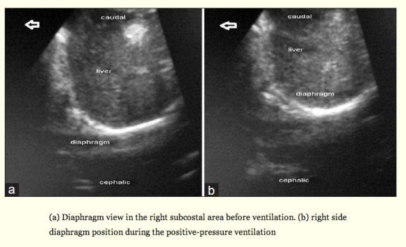
SonoStudy: Right subcostal diaphragm view to confirm ETT placement #FOAMed

In the Apr 2013 issue of Int J Crit Illn Inj Science, the authors from Iran attempt to identify the sensitivity, specificity, positive predictive value and accuracy of the right subcostal diaphragm view in the immediately paralyzed and intubated patient to confirm endotracheal intubation (versus esophageal intubation). This study comes from prior studies done suggesting that by visualizing diaphragm movement with ventilation or BVM, you can ascertain that the ET tube was placed in the trachea and not the esophagus. This adds to the growing number of studies that also discusses visualization of the lung sliding sign after intubation to confirm endotracheal intubation, and the ease at which you can identify the tube in the trachea and the esophagus upon trans-jugular notch view and identification of the ring down artifact through the trachea or a tube shadow beyond the esophagus, respectively.

To view a prior post on airway US and ETT placement confirmation, go here. For a fun airway guided talk by Ultrasound Podcast on it all, go here, and here.

So, after you intubate – look over the jugular notch for the ring down artifact through the trachea with the absence of esophageal shadow, then look for lung sliding on both sides of the chest with BVM or ventilation (this can also assess for pneumothorax prior to intubation), then look at the diaphragm for movement with ventilation post intubation. None will show you exactly where the end of the ET tube is, but you’ll know that youre not in the esophagus with good confidence – especially if your capnography isnt accurate due to post- cardiac arrest and your Xray machine/tech is busy or in the bathroom.

The abstract is below:

Aims:

To assess the sensitivity and specificity of right subcostal ultrasound view to confirm correct endotracheal tube intubation (ETT).

Materials and Methods:

In this prospective study, apneic or paralyzed patients who had an indication of intubation were selected. Intubation and ventilation with bag were performed by the skilled third-year emergency medicine residents. The residents, following a brief training course of ultrasonography, interpreted the diaphragm motion, and identified either esophageal or tracheal intubation. The confirmation of ETT placement was done by the sonographer. Sensitivity, specificity, positive predictive value, negative predictive value, and accuracy were calculated for tracheal versus esophageal intubation.

Results:

A total of 57 patients aged 59 ± 5 who underwent ETT insertion were studied. Thirty-four of them were male (60%). Ultrasound correctly identified 11 out of 12 esophageal intubations for a sensitivity of 92% (95% CI = 62-100), but misidentified one esophageal intubation as tracheal. Sonographers correctly identified 43 out of 45 (96%) tracheal intubations for a specificity of 96% (95% CI = 85-99), but misdiagnosed two tracheal intubations as esophageal.

Conclusions:

This study suggests that diaphragm motion in right subcostal ultrasound view is an effective adjunct to diagnose ETT place in patients undergoing intubation in emergency department.

Likely the best description from the article is below:”…Studies have shown that ultrasound can diagnose paralyzed diaphragm.[16,17] Therefore, it seems practical to detect diaphragm motion during the positive-pressure ventilation by using ultrasound imaging of it. Hsieh, et al. studied the use of diaphragm motion for confirmation of ETT correct placement on 59 children in prenatal intensive care unit (PICU) setting. They diagnosed all intubations correctly with ultrasound imaging with sensitivity and specificity of 100%; however, they had only two esophageal intubations.[25] Kerrey, et al.[26] used diaphragm motion view of ultrasound in confirmation of correct ETT placement in 66 children in PICU. They evaluated the diagnosis of right main bronchus intubation. In their study, the sensitivity and specificity were 88% and 64%, respectively. Although sensitivity and specificity of the ultrasound in our study were high, we cannot strongly state that the ultrasound is a sensitive and specific method, because the confidence interval for sensitivity and specificity were rather wide. Studies have identified the intubation of the right main bronchus using diaphragm motion or observation of the lung sliding sign by placing the probe in the left side of the chest; however, left side of the chest is not as good as the right side for seeing the diaphragm motion.[22,26] If the patient is paralyzed or apneic, it would be suitable to evaluate the diaphragm motion in the right side of the chest to diagnose the esophageal intubation.”

Screen Shot 2014-04-02 at 10.15.58 AM

SonoStudy: Ultrasound of heart and lungs identify preeclamptic pregnant patients at risk

In anesthesiology literature (not yet on pubmed), heart and lung ultrasound has entered the mix to identify pregnant patients at risk for respiratory failure – looking for hypocontractile heart and B lines! Oh yeah! It has received a lot of press, see here, here and here :

“Chicago — (March 18, 2014)

An ultrasound of the lungs could help doctors quickly determine if a pregnant woman with preeclampsia is at risk for respiratory failure, suggests preliminary research published in the April issue of Anesthesiology.

About 60,000 women worldwide die as a result of preeclampsia, which causes severely high blood pressure. Potential complications include stroke, bleeding and excess fluid in the lungs – called pulmonary edema – which can lead to respiratory failure.  The study suggests a lung ultrasound can help doctors easily learn whether a woman with preeclampsia is suffering from pulmonary edema and ensure she receives the correct treatment.

“Lung Ultrasound is fast, safe, noninvasive and easy to use,” said Marc Leone, M.D., Ph.D., lead author of the study and vice chair of the department of anesthesiology and critical care medicine, Hopital Nord, Marseille, France. “We found it allowed us to quickly assess whether a woman with preeclampsia had pulmonary edema and confirm the severity of the condition.”

Doctors often measure urine output to determine if a woman needs fluid administration, but the results are wrong about half of the time. “Lung ultrasound enables the medical team to identify which women really need the fluid treatment,” noted Dr. Zieleskiewicz, the study’s  first author.

Pulmonary edema is typically caused by heart failure, but also can be caused by lung inflammation. Researchers analyzed the use of lung ultrasounds, which can assess lung  edema, are easier to use than cardiac ultrasound and can be performed with devices commonly found in maternity wards. Lung ultrasound highlights white lines mimicking comet tails, irradiating from the border of the lungs. These lines are the reflection of water in the lungs.  The detection of three or more lines strongly suggests the diagnosis of pulmonary edema.

Researchers performed both cardiac and lung ultrasounds before and after delivery in 20 women with severe preeclampsia.  Five of the 20 women (25 percent) had pulmonary edema prior to delivery according to lung ultrasound, while four (20 percent) had the condition according to the cardiac ultrasound. The lung ultrasound identified a patient with non-cardiac pulmonary edema, which the cardiac ultrasound did not detect.

The test results could help ensure that pregnant women with pulmonary edema not be given intravenous or excess fluids, which worsens the condition and can lead to respiratory failure.  Typically, women with pulmonary edema are treated with oxygen and medication to lower the blood pressure or rid the body of excess fluid.  In real time, lung ultrasound also serves to observe improvement or worsening of pulmonary edema.”