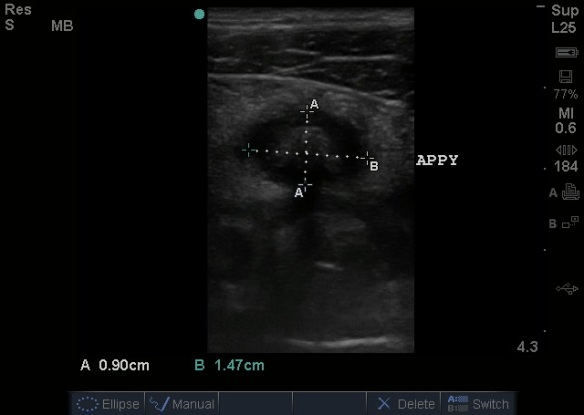
In a study published in AJR, a very hot topic was reviewed. 2 centers. 160 kids. Ultrasound and appendectomy with comparison to operative report. Do I have your attention now? This is a tough one, ultrasound for appendicitis is being recommended by pediatricians, radiologists, emergency physicians and surgeons. A big limitation was thought that ultrasound is not great for differentiating perforated from non-perforated appendicitis…. in addition to other limitations including bowel gas scatter limiting view of the entire appendix, and variations in appendix size that may have a false positive for appendicitis if diameter size alone is used as the indicator. Well, it isnt perfect – we know that.
Now, to review, appendicitis is diagnosed by applying the linear (or curvilinear if added depth is needed) probe to the area where the patient points to noting maximal pain, with the indicator toward the patient’s right side. Graded compression is then performed in that region which should displace and flatten bowel, identifying the psoas muscle and the transverse view of the iliac vessels. The appendix usually is located just anterior to these structures coming off of the cecum, and is normally compressible without being more than 6mm in diameter. It may be in its transverse or longitudinal view depending on anatomy. The entire appendix should be viewed, including to its tip. Be sure to view it in two orthogonal planes (rotate probe 90 degrees) to ensure it is the appendix, as a lymph node may look very similar to a transverse appendix but will not elongate into a tubular structure when viewed in its longitudinal plane. Here are some views of a positive appendicitis (absence of compressibility with attempts, dilated appendix):
Appendicitis by Ultrasound: A greater than 6mm in diameter, aperistaltic, non-compressible appendix +/- appendecolith.
Ultrasound Podcast posted a great video a year ago on the “how-to” for appendix ultrasound and why to go to ultrasound first in the work up of appendicitis:
Let’s go back to the study:
“OBJECTIVE. Acute appendicitis is the most common condition requiring emergency surgery in children. Differentiation of perforated from nonperforated appendicitis is important because perforated appendicitis may initially be managed conservatively whereas nonperforated appendicitis requires immediate surgical intervention. CT has been proved effective in identifying appendiceal perforation. The purpose of this study was to determine whether perforated and nonperforated appendicitis in children can be similarly differentiated with ultrasound.
MATERIALS AND METHODS. This retrospective study included 161 consecutively registered children from two centers who had acute appendicitis and had undergone ultra-sound and appendectomy. Ultrasound images were reviewed for appendiceal size, appearance of the appendiceal wall, changes in periappendiceal fat, and presence of free fluid, abscess, or appendicolith. The surgical report served as the reference standard for determining whether perforation was present. The specificity and sensitivity of each ultrasound finding were determined, and binary models were generated.
RESULTS. The patients included were 94 boys and 67 girls (age range, 1-20 years; mean, 11 ± 4.4 [SD] years) The appendiceal perforation rate was significantly higher in children younger than 8 years (62.5%) compared with older children (29.5%). Sonographic findings associated with perforation included abscess (sensitivity, 36.2%; specificity, 99%), loss of the echogenic submucosal layer of the appendix in a child younger than 8 years (sensitivity, 100%; specificity, 72.7%), and presence of an appendicolith in a child younger than 8 years (sensitivity, 68.4%; specificity, 91.7%).
CONCLUSION. Ultrasound is effective for differentiation of perforated from nonperforated appendicitis in children.”
Interestingly, a multi-organizational group came together for guidelines published in a study in Pediatric Emergency Care. : abstract below:
“The objective of this study was to compare usage of computed tomography (CT) scan for evaluation of appendicitis in a children’s hospital emergency department before and after implementation of a clinical practice guideline focused on early surgical consultation before obtaining advanced imaging.
METHODS:
A multidisciplinary team met to create a pathway to formalize the evaluation of pediatric patients with abdominal pain. Computed tomography scan utilization rates were studied before and after pathway implementation.
RESULTS:
Among patients who had appendectomy in the year before implementation (n = 70), 90% had CT scans, 6.9% had ultrasound, and 5.7% had no imaging. The negative appendectomy rate before implementation was 5.7%. In patients undergoing appendectomy in the postimplementation cohort (n = 96), 48% underwent CT, 39.6% underwent ultrasound, and 15.6% had no imaging. The negative appendectomy rate was 5.2%. We demonstrated a 41% decrease in CT use for patients undergoing appendectomy at our institution without an increase in the negative appendectomy rate or missed appendectomy. The results were even more striking when comparing the rate of CT scan use in the subset of patients undergoing appendectomy without imaging from an outside hospital. In these patients, CT scan utilization decreased from 82% to 20%, a 76% reduction in CT use in our facility after protocol implementation.
CONCLUSIONS:
Implementation of a clinical evaluation pathway emphasizing examination, early surgeon involvement, and utilization of ultrasound as the initial imaging modality for evaluation of abdominal pain concerning for appendicitis resulted in a marked decrease in the reliance on CT scanning without loss of diagnostic accuracy.”
Why talk about this? Well, there is ALWAYS, always, ALWAYS press about how ultrasound can and should be used for appendicitis evaluation in pediatric patient for radiation exposure minimization. It does have false negatives and false positives though – as with all thing ultrasound, you must know it’s strengths and weaknesses….and correlate clinically 🙂


SIR NICE STUDY RELATED TO APPENDIX , I AM SENIOR CONSULTANT RADIOLOGIST ,PRESENTLY WORKING AS ASSISTANT PROFESSOR IN RADIOLOGY DEPARTMENT IN
NCR REGION , INDIA
Thank you Dr. Vijay!